
-
-
SHOP

Multiplex Immunohistochemistry: Chromogenic vs. Fluorescent Techniques
![]() 2025-05-08
2025-05-08
By admin
Multiplex immunohistochemistry (mIHC) is a strong tool in medical studies. It lets scientists see many markers on one tissue piece. This method is key for exploring complex body processes, like cancer environments, immune cell actions, and disease growth. Two main detection ways lead mIHC: chromogenic mIHC and fluorescent mIHC. Each has special strengths and weaknesses, making them fit for certain cases. This blog dives into these methods, comparing their uses, benefits, and challenges. It helps researchers pick the best approach for their work.
What Is Multiplex Immunohistochemistry
Multiplex immunohistochemistry allows spotting several protein markers on one tissue slice at once. It saves valuable samples. It also shows how markers relate in space. Unlike old singleplex IHC, which marks one thing per slide, mIHC gives more data. This makes it great for research, clinical tests, and cancer studies.
The choice between chromogenic mIHC and fluorescent mIHC depends on study goals, tools available, sample kind, and the need for number-based or quality-based results. Below, we explore each method’s details, showing their pros and cons in real cases.
Chromogenic mIHC: Principles and Applications
How Chromogenic mIHC Works
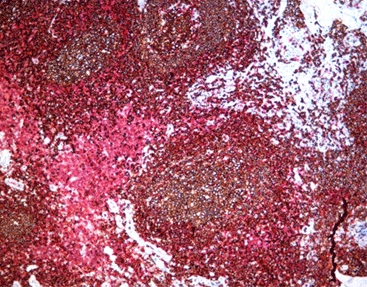
Chromogenic mIHC uses enzyme-driven reactions. These often involve horseradish peroxidase (HRP) or alkaline phosphatase (AP). They trigger colored deposits at the marker spot. These deposits, like 3,3’-diaminobenzidine (DAB) or clear chromogens, show up under a regular light microscope. Improved methods, like tyramide signal amplification (TSA), boost sensitivity. They help find low-amount proteins.
Advantages of Chromogenic mIHC
- Affordable and Easy to Use: Chromogenic mIHC works with standard brightfield microscopes. These are common in labs, so no special tools are needed.
- Lasting Marks: The colored deposits stay stable. Slides can be kept for years without losing clarity, unlike fluorescent dyes that fade.
- Familiar Process: Chromogenic mIHC fits with usual lab routines. This makes it simple to use in medical settings.
- Clear Tissue View: Hematoxylin staining shows tissue structure well. It helps with shape assessment.
Limitations of Chromogenic mIHC
- Few Markers at Once: Chromogens have wide color ranges. Only 3–5 markers can be seen clearly without mixing.
- Basic Number Analysis: Chromogen strength isn’t very exact. This makes precise counting harder than with fluorescent methods.
- Color Mixing: Overlapping chromogenic marks can blend. This makes it tough to study markers in the same spot.
Best Scenarios for Chromogenic mIHC
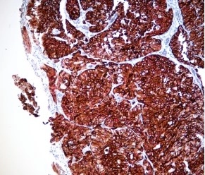
Chromogenic mIHC shines in medical tests where preserved tissues are common. It’s also good for quick, budget-friendly analysis. For example, in lung cancer tests, chromogenic mIHC panels (e.g., TTF1, p40, PD-L1, CD8) can show tumor types and key markers on one slide. This saves tissue for other tests.
Fluorescent mIHC: Principles and Applications
How Fluorescent mIHC Works
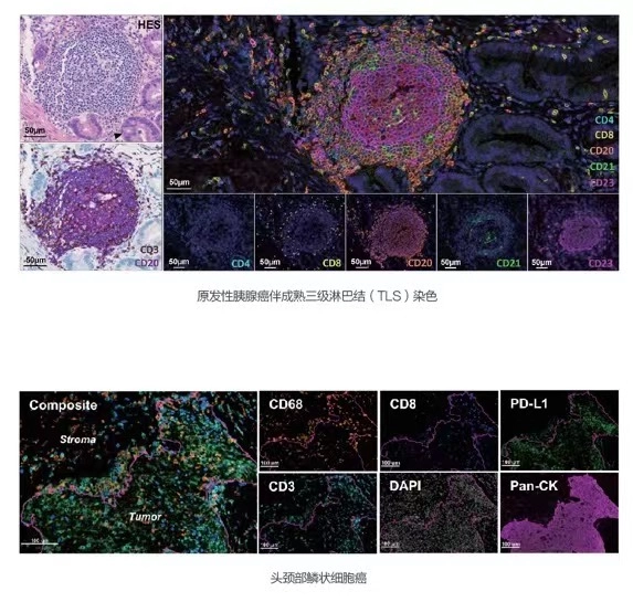
Fluorescent mIHC uses glowing tags linked to antibodies or TSA. These tags shine at specific light wavelengths when activated. Multispectral imaging and signal separation spot multiple glowing tags, even if their signals overlap. This method needs fluorescence microscopes or slide scanners with the right filters.
Advantages of Fluorescent mIHC
- Many Markers at Once: Fluorescent mIHC can spot 5–10+ markers on one slide. It’s perfect for complex studies like cancer immune mapping.
- Exact Number Analysis: Glowing tags give a wide, steady signal range. This allows accurate counting of protein levels at the cell level.
- Better Marker Overlap Study: Signal separation avoids mixing. This makes fluorescent mIHCgreat for studying proteins in the same cell area.
- Stronger Sensitivity with TSA: TSA boosts glow signals. It finds low-amount targets clearly.
Limitations of Fluorescent mIHC
- Costly Tools: Fluorescent mIHC needs special fluorescence microscopes or multispectral systems. These can be pricey.
- Fading Signals: Glowing tags lose strength over time. Careful storage and imaging are needed.
- Natural Glow Issues: Preserved tissues, like spleen or kidney, may glow on their own. This can confuse signals. Multispectral imaging helps, but it’s complex.
Best Scenarios for Fluorescent mIHC
Fluorescent mIHC is best for research needing many markers and exact counts. It’s great for studying cancer immune settings. For example, in breast cancer work, fluorescent mIHC can map CD8+ T cells, TFH cells, and B cells in lymphoid structures. This shows spatial links vital for immunotherapy success.
Comparing Chromogenic and Fluorescent mIHC: A Practical Guide
To help researchers choose between chromogenic mIHC and fluorescent mIHC, this table sums up key differences:
|
Feature |
Chromogenic mIHC |
Fluorescent mIHC |
|
Marker Count |
3–5 markers |
5–10+ markers |
|
Tools |
Standard brightfield microscope |
Fluorescence microscope or scanner |
|
Signal Durability |
Permanent, no fading |
Fades over time |
|
Number Analysis |
Basic counting |
Highly exact counting |
|
Marker Overlap |
Limited due to mixing |
Clear with signal separation |
|
Cost |
Cheaper, widely available |
More expensive, special tools |
|
Best Use |
Medical tests, preserved tissues, routine work |
Research, immune studies, many markers, IHC Reagents |
When to Choose Chromogenic mIHC
- Routine Lab Work: It fits existing lab processes with standard tools.
- Tight Budget: It’s good for labs without fluorescence tools.
- Long Storage: Slides stay clear without signal loss.
When to Choose Fluorescent mIHC
- Complex Studies: It suits work needing many markers and cell-level analysis.
- Overlap Studies: It’s ideal for exact marker co-location data.
- Number Needs: It gives accurate marker counts for research.
Celnovte: Your Trusted mIHC Supplier
For researchers seeking top-quality mIHC tools, Celnovte is a leading provider. Celnovte focuses on advanced IHC and mIHC supplies. They offer items like the Multiplex Immunohistochemical (mIHC) Kit and Immune Chromogenic Reagent Double Stain I. Their cutting-edge reagents, including TSA-based chromogenic and fluorescent systems, deliver dependable, clear results. With a focus on quality and new ideas, Celnovte helps researchers achieve accurate, repeatable mIHC outcomes. They are a trusted partner for both chromogenic mIHC and fluorescent mIHC work, Celnovte Homepage.
FAQs About Chromogenic and Fluorescent mIHC
Q1. What’s the key difference between chromogenic mIHC and fluorescent mIHC?
A1. Chromogenic mIHC uses enzyme-based color deposits. They show under brightfield microscopes. In contrast, fluorescent mIHC uses glowing tags seen with fluorescence microscopes. Chromogenic is more lasting and cheaper. Fluorescent offers more markers and exact counts.
Q2. How many markers can chromogenic mIHC spot compared to fluorescent mIHC?
A2. Chromogenic mIHC usually spots 3–5 markers. Color overlap limits it. However, fluorescent mIHC can spot 5–10 or more. Signal separation and multispectral imaging make this possible.
Q3. Is fluorescent mIHC good for preserved tissues?
A3. Yes, fluorescent mIHC works well for preserved tissues. But natural glow can be a problem. Multispectral imaging and signal separation reduce this issue. They ensure clear results.
Q4. Which method is better for marker overlap studies?
A4. Fluorescent mIHC is better for overlap studies. It separates mixed signals clearly. This makes it ideal for studying proteins in the same cell spot.
Take Your mIHC Research to the Next Level
Whether you’re doing routine medical tests or advanced cancer research, picking the right mIHC method matters. Chromogenic mIHC is easy to use and stable. Fluorescent mIHC offers unmatched marker counts and exact data. Check out Celnovte’s mIHC tools, like the Immune Chromogenic Reagent Double Stain II. Ready to improve your studies? Contact Celnovte today to explore your mIHC needs and unlock your tissue samples’ full potential.


