
-
-
SHOP

New Product – Fast Frozen IHC solution
Celnovte takes pride in presenting an advanced Fast Frozen Kit, featuring improved fixative formulation and optimized tissue fixation methods. This innovation ensures the consistency of frozen tissue morphology with paraffin-embedded tissues, preserving tissue integrity without compromising structure. The result is enhanced tissue morphology, leading to crisper and clearer staining—an invaluable aid for pathologists in their result interpretation.
Section 1: Tissue Section Preparation
Prepare fresh tissue samples of appropriate size and place tissue chunks on a support with an adequate amount of OTC embedding medium. After rapid freezing of the tissue in a temperature-controlled cryostat (commonly between -21°C to -26°C), cut the frozen tissue blocks into thin slices of approximately 5μm thickness. Leave the tissue slices slightly on the block, then gently press a labeled room temperature slide onto the tissue, allowing the tissue to quickly melt onto the glass slide.
Note: Different tissues may have different optimal freezing temperatures.
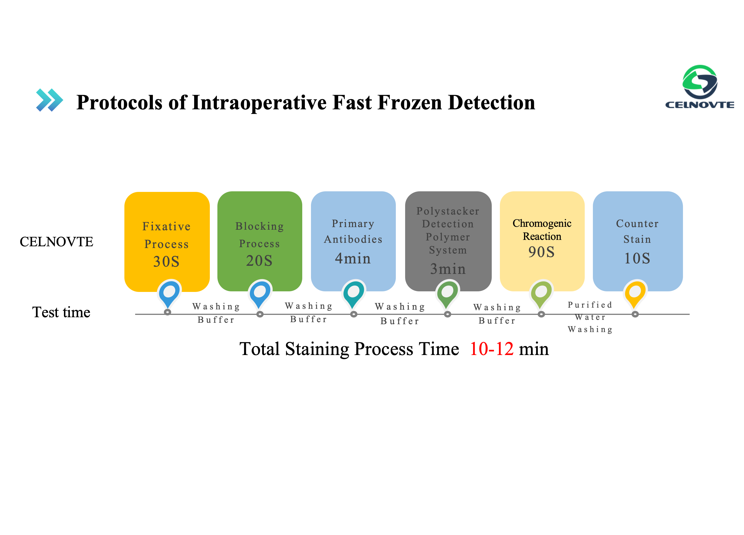
Section 2: Staining Process
2.1 Fixation of Frozen Section Tissues
Immediately place the adhered frozen tissue slides into a fixation solution and fix for 30 seconds to 1 minute. Note: For optimal staining results, use the fixation solution provided by Sanotec. Other brand fixatives should be validated by pathology technicians for effectiveness before use.*
2.2 Post-Fixation Rinse
After fixing the slides, remove them and gently shake them in tap water to completely remove the fixation solution and OTC embedding medium from the slides. Note: Rinse gently to prevent tissue detachment.
2.3 Blocking
After removing the residual fixation solution and embedding medium, use an immunohistochemistry pen to delineate the area of the tested tissue. Add an appropriate amount of blocking reagent and incubate for 20 seconds. Then wash the slides with TBS washing solution for 30 seconds.
2.4 Add Primary Antibody*
Remove excess washing solution from the slides, add an appropriate amount of Sanotec frozen immunohistochemistry-specific primary antibody reagent within the delineated area, and incubate for 2-4 minutes. Wash the slides with TBS washing solution for 30 seconds. Note: Ensure the antibody is at room temperature before use. Increasing the antibody incubation temperature can enhance staining.
2.5 Add Secondary Antibody
Remove excess washing solution from the slides, add an appropriate amount of Sanotec frozen immunohistochemistry-specific secondary antibody reagent within the delineated area, and incubate for 2-3 minutes. Wash the slides with TBS washing solution for 30 seconds. Note: Do not excessively shorten the washing time to avoid non-specific background staining.
2.6 Add DAB
Remove excess washing solution from the slides, add an appropriate amount of DAB chromogenic solution within the delineated area, and incubate for about 90 seconds. Then rinse with pure water for 30 seconds. Note: Freshly prepare the DAB solution before use. The effective duration of the prepared DAB solution is 6 hours at room temperature (25°C) and 12 hours at 4°C.
2.7 Counterstaining with Hematoxylin
Add an appropriate amount of hematoxylin counterstaining solution, incubate for about 10 seconds, and then rinse with pure water for 30 seconds. Note: The counterstaining solution in this kit has strong staining ability. Do not overextend the counterstaining time, especially for nuclear staining, as prolonged counterstaining may affect the interpretation of positive results.
2.8 Dehydration and Mounting
Section 3: Staining Results
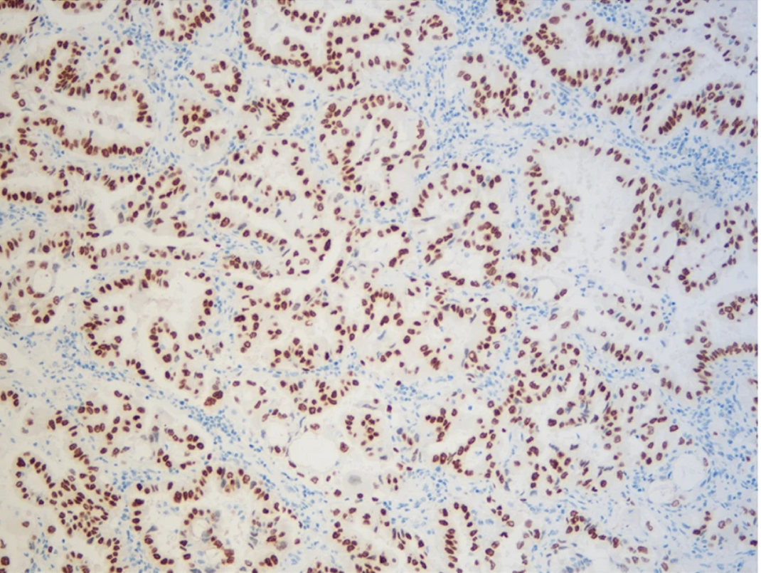
TTF-1


