 PRODUCT CATEGORY
CONTACT US
Phone
Email
Address
6 Cuizhu St, Zhong Yuan Qu, Zheng Zhou Shi, He Nan Sheng, China, 450001
Multiplex Immunofluorescence(mIF) Kit
In stock in USA.
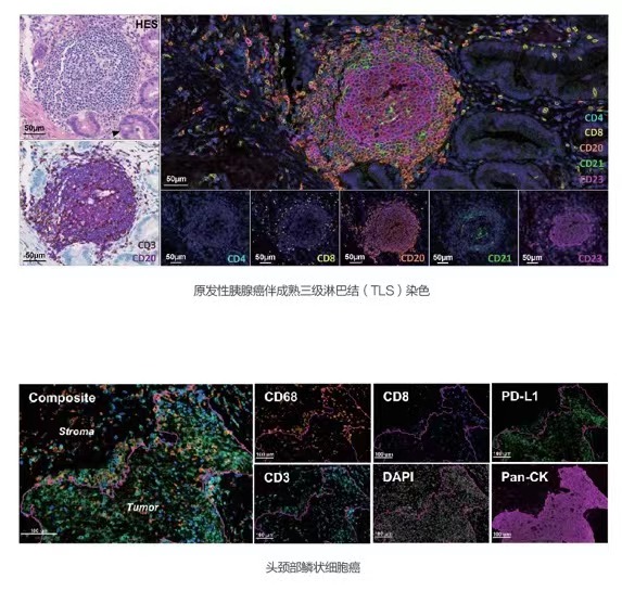
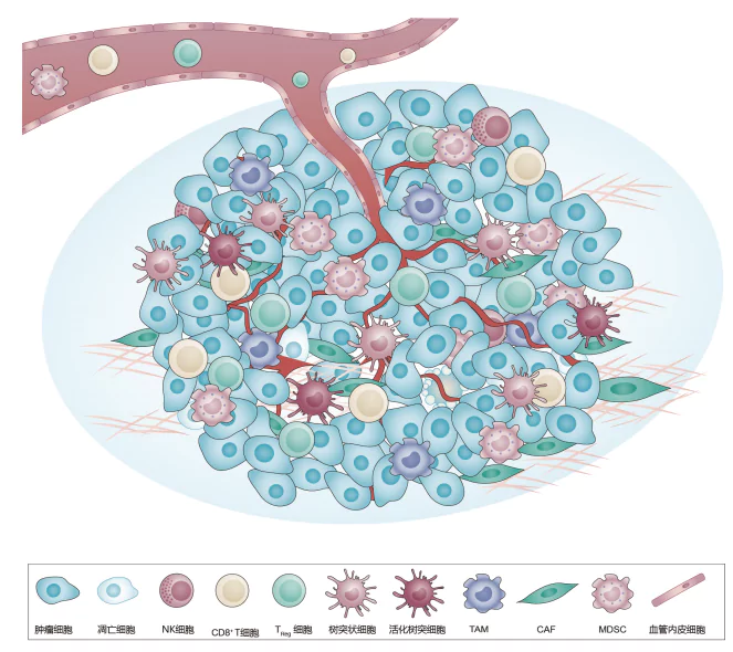
The Multiplex Immunofluorescence(mIF) technology allows for the simultaneous labeling of multiple biomarkers on a single tissue section, enabling comprehensive analysis of the tumor immune microenvironment, quantitative evaluation of cell phenotypes, and detailed understanding of cell interactions and spatial relationships. This is achieved using tyramide signal amplification (TSA) technology with horseradish peroxidase (HRP) and a proprietary Microstacker Polymer.
Shop online: https://shop.celnovte.com/collections/multiplex-ihc
Product Features
[Intended Use]
Research Use only
Multiplex Immunofluorescence (mIF) is an advanced technique widely used in pathology and biomedical research to simultaneously detect and visualize multiple biomarkers within a single tissue section. It enables detailed analysis of protein expression, interactions, and spatial distribution within the tissue microenvironment.
– Allows protocol runs onboard with CNT320/330/360
– 4-8 markers detection simultaneously
– No species restrictions
– High Sensitivity and Specificity
[Specifications]
| Cat# | Product name | Format | Components | Shop online |
| FM2007 | 7-color multiplex fluorescence IHC detection kit | 20T / 50T | Endogenous Peroxidase Blocking reagent Anti-Mouse/Rabbit HRP-Polymer TSA Fluorophore CM480 (Ex:425nm, Em:485nm) TSA Fluorophore CM520 (Ex:490nm, Em:520nm) TSA Fluorophore CM570 (Ex:550nm, Em:570nm) TSA Fluorophore CM620 (Ex:585nm, Em:615nm) TSA Fluorophore CM690 (Ex:675nm, Em:690nm) TSAFluorophore CM780 (Ex:750nm, Em:770nm) Spectral DAPI concentrate (Ex:355nm, Em:460nm) TSA-biotin Amplification Diluent PBS Buffer |
🛒 Buy now |
| FM2004 | 4-color multiplex fluorescence IHC detection kit | 20T / 50T | Endogenous Peroxidase Blocking reagent Anti-Mouse/Rabbit HRP-Polymer TSA Fluorophore CM520 (Ex:490nm, Em:520nm) TSA Fluorophore CM570 (Ex:550nm, Em:570nm) TSA Fluorophore CM690 (Ex:675nm, Em:690nm) Spectral DAPI concentrate (Ex:355nm, Em:460nm) Amplification Diluent |
🛒 Buy now |
| FM2017 | 7-color multiplex fluorescence IHC detection kit (rabbit-on-rodent) | 20T / 50T | Endogenous Peroxidase Blocking reagent Anti-Rabbit HRP-Polymer TSA Fluorophore CM480 (Ex:425nm, Em:485nm) TSA Fluorophore CM520 (Ex:490nm, Em:520nm) TSA Fluorophore CM570 (Ex:550nm, Em:570nm) TSA Fluorophore CM620 (Ex:585nm, Em:615nm) TSA Fluorophore CM690 (Ex:675nm, Em:690nm) TSAFluorophore CM780 (Ex:750nm, Em:770nm) Spectral DAPI concentrate (Ex:355nm, Em:460nm) TSA-biotin Amplification Diluent PBS Buffer |
🛒 Buy now |
| FM2014 | 4-color multiplex fluorescence IHC detection kit (rabbit-on-rodent) | 20T / 50T | Endogenous Peroxidase Blocking reagent Anti-Rabbit HRP-Polymer TSA Fluorophore CM520 (Ex:490nm, Em:520nm) TSA Fluorophore CM570 (Ex:550nm, Em:570nm) TSA Fluorophore CM690 (Ex:675nm, Em:690nm) Spectral DAPI concentrate (Ex:355nm, Em:460nm) Amplification Diluent |
🛒 Buy now |
| FT480 | TSA Fluorophore CM480 | 20T / 50T | TSA Fluorophore CM480 (Ex:425nm, Em:485nm), Amplification Diluent |
🛒 Buy now |
| FT520 | TSA Fluorophore CM520 | 20T / 50T | TSA Fluorophore CM520 (Ex:490nm, Em:520nm), Amplification Diluent |
🛒 Buy now |
| FT570 | TSA Fluorophore CM570 | 20T / 50T | TSA Fluorophore CM570 (Ex:550nm, Em:570nm), Amplification Diluent |
🛒 Buy now |
| FT620 | TSA Fluorophore CM620 | 20T / 50T | TSA Fluorophore CM620 (Ex:585nm, Em:615nm), Amplification Diluent |
🛒 Buy now |
| FT690 | TSA Fluorophore CM690 | 20T / 50T | TSA Fluorophore CM690 (Ex:675nm, Em:690nm), Amplification Diluent |
🛒 Buy now |
| FT780 | TSA Fluorophore CM780 | 20T / 50T | TSAFluorophore CM780 (Ex:750nm, Em:770nm) Amplification Diluent TSA-biotin PBS buffer |
🛒 Buy now |
| FTDAPI | Spectral DAPI concentrate | 20T / 50T | Spectral DAPI concentrate (Ex:355nm, Em:460nm) |
🛒 Buy now |
[Documentation]
| Brochure | ↕️ Download |
| User Guide | ↕️ Download |


![[Manual] MicroStacker™ Polymer Detection Kit Universal, 1 Step [Manual] MicroStacker™ Polymer Detection Kit Universal, 1 Step](https://www.celnovte.com/wp-content/uploads/2020/08/123-3.jpg)
![[RX] MicroStacker™ Rabbit-on-Rodent HRP-Polymer [RX] MicroStacker™ Rabbit-on-Rodent HRP-Polymer](https://www.celnovte.com/wp-content/uploads/2020/08/4-1.gif)
![[Manual] MicroStacker™ Polymer Detection Kit, Enhanced, 2-Step [Manual] MicroStacker™ Polymer Detection Kit, Enhanced, 2-Step](https://www.celnovte.com/wp-content/uploads/2020/08/1-1.gif)