
-
-
SHOP

Why IHC Is Classified as an Immunoassay
![]() 2025-12-26
2025-12-26
By admin
Introduction: Unlocking the Molecular Blueprint of Disease
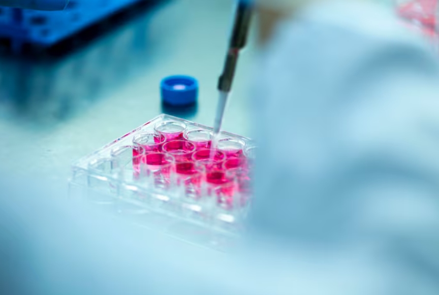
Walk into any busy pathology lab, and you’ll see IHC slides everywhere. Brown stains on breast tumors, red and blue on lymphoma nodes, and nuclear dots on colon biopsies. Those colors aren’t just pretty—they’re the direct result of an immune reaction happening right on the glass slide. At its core, IHC is nothing more (and nothing less) than an immunoassay that happens inside real tissue instead of a test tube. That single fact explains both its power and why it’s grouped with ELISA, lateral flow tests, and Western blots under the big “immunoassay” umbrella.
This plain-language piece explains exactly why IHC earns that label, how the antibody-antigen handshake drives every step, and how companies like Celnovte keep pushing the technology forward with sharper reagents and smarter machines.
The Foundational Link: What Type of Assay is IHC?
Defining Immunohistochemistry (IHC)
IHC combines two worlds: histology (looking at tissue structure) and immunology (using antibodies to grab specific targets). You fix a thin slice of tissue on a slide, add an antibody that only likes one particular protein, let it bind, and then make that binding visible with color. Simple in theory, incredibly useful in practice.
The Core Mechanism: Antibody-Antigen Recognition
Everything rides on one biological trick—the antibody sticking tightly and exclusively to its matching antigen. That lock-and-key fit is the same reaction that powers every immunoassay on the planet.
In IHC, the “analyte” is the protein you care about (say CD8 on T-cells or MUC-5AC in gastric cancer). The “detector” is a carefully chosen antibody. Because the test depends 100 % on that immune recognition event, IHC is, by textbook definition, an immunoassay—only it shows you not just “how much” but “exactly where”.
Immunoassays in Context: Bridging Techniques
Most immunoassays work on blood or urine and give you a number. IHC works on solid tissue and gives you a picture. That picture is what makes it priceless in surgical pathology—pathologists can see whether HER2 sits on the membrane, whether PD-L1 coats the tumor cells, or whether MMR proteins have vanished from the nuclei.
Defining Principles of Immunodiagnostics
Specificity and Sensitivity: The Role of Antibodies
A sloppy antibody ruins the whole test. Celnovte builds its own clones from scratch—more than 130 monoclonal (MMab) and recombinant (RMab) primary antibodies—so lot-to-lot performance stays tight. Independent proof comes from NordiQC runs: 47 of those in-house antibodies have scored “optimal” or “good”, meaning labs worldwide trust them for clean, crisp staining on real patient samples.
i
Signal Amplification and Visualization
Many diagnostic proteins are present in tiny amounts. You need a detection system that turns a faint whisper into a loud, clear signal without shouting nonsense in the background.
Celnovte’s Advanced Solutions for Precision IHC
Innovating Detection: The MicroStacker™ Detection System
That’s exactly what the MicroStacker™ Detection System does. Using a patented micro-polymer stacking design (micropolymer scaffold, HRP layering, Fab’ labeling, and conjugation chemistry), it packs many more enzyme molecules onto each antibody than old bulky dextran polymers ever could. Result: much stronger brown color on weak markers. And because it skips biotin and streptavidin completely, tissues full of natural biotin—liver, kidney, brain—stay snow-white instead of turning dirty brown.
Standardization in Clinical Pathology
Hand-staining twenty slides the same way is tough. One tech presses the wash bottle a little longer, another lets the chromogen sit thirty seconds extra—suddenly the stains don’t match.
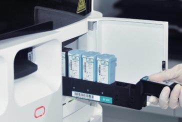
Streamlining Workflow: The CNT300 Full Automatic Multiplex IHC Stainer
The CNT300 Full Automatic Multiplex IHC Stainer removes that human wiggle. It runs the entire protocol—deparaffinization to counterstain—exactly the same way every time, slide after slide. Since 2018, Celnovte has placed over 800 automated IHC platforms worldwide. Labs report fewer repeats, faster sign-out, and happier pathologists.
Clinical Impact and Future Directions
IHC in Cancer Diagnostics and Companion Diagnostics
Today’s oncology leans heavily on IHC immunoassays. Celnovte’s MMR panel (MLH1, MSH2, MSH6, PMS2) is run thousands of times a week to spot Lynch syndrome or predict immunotherapy response. The same goes for ADC companion diagnostics—clear HER2 membrane staining or strong TROP2 expression decides whether a patient gets an expensive new drug conjugate.
Ensuring Reliability: High-Quality Primary Antibodies
Celnovte backs every antibody with full regulatory compliance: ISO13485, ISO9001, NMPA, GMP, FDA registration, and CE IVDR. That paperwork means the bottle you open in Seoul works exactly like the one opened in São Paulo.
The Future Landscape of Immunoassay Technology
Speed is the next frontier. Celnovte already offers Fast Frozen IHC solutions that drop turnaround time for intraoperative slides to around 10 minutes with no loss of confidence. Surgeons no longer close the patient and hope—they get the IHC answer while the case is still open.
Conclusion: Elevating Precision in Pathology
IHC earns its immunoassay badge because every brown dot on the slide comes from an antibody hugging its antigen—pure immunology at work. Companies like Celnovte keep raising the bar with cleaner detection (MicroStacker™), tighter antibodies (NordiQC-proven clones), and bullet-proof automation (CNT300). The result? Pathologists sign out tougher cases faster, oncologists pick better drugs, and patients get the right treatment the first time.
FAQ
Q: Why is IHC considered a morphological immunoassay?
A: It uses classic antibody-antigen binding (immuno) but shows the result inside the actual tissue structure (morphological), telling you not just “present or not” but exactly where the protein lives.
Q: What is the main advantage of the MicroStacker™ system?
A: Patented micro-polymer stacking gives far higher sensitivity and completely skips biotin-related background problems.
Q: How does Celnovte ensure the quality of its primary antibodies?
A: We clone and manufacture them in-house; 47 have earned optimal or good scores in independent NordiQC assessments.
Q: What are the benefits of using an automated IHC stainer like the CNT300?
A: It removes hand-staining variability, cuts processing time, and delivers consistent, clean slides run after run—exactly what busy clinical labs need.