
-
-
SHOP

What Ki-67 Marker Reveals About HPV-Related Cell Proliferation
![]() 2025-11-20
2025-11-20
By admin

Understanding the Ki-67 Marker in HPV Context
What Is Ki-67 and Why Does It Matter
Ki-67 acts as a protein found inside the cell nucleus that helps manage how cells divide. You can spot it only when cells are busy growing—during the G1, S, G2, and mitosis stages—but it vanishes completely in cells that are taking a break in the G0 phase. This clear on-off switch turns Ki-67 into a handy sign for checking how actively cells multiply in tissue samples.
Doctors pay close attention to Ki-67 in cancer checks because lots of it usually means cells are dividing too fast, a red flag for unusual growth or even tumors. When HPV steps in, the virus messes with the normal brakes on cell division. Watching Ki-67 levels then gives clues about whether tissue might be heading toward abnormal changes or full-blown cancer.
Role of Ki-67 in HPV-Infected Cells
HPV pushes cells to make too much of its troublemaking proteins, E6 and E7. These proteins knock out the cell’s own safety controls, so division spins out of control—a key step toward cancer. Ki-67 steps up here by pointing out spots where this wild growth happens because of the virus.
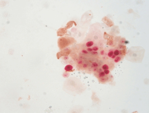
In healthy cervical lining, Ki-67 shows up just in the bottom layer of cells. But once HPV hits or tissue turns dysplastic, you see Ki-67-positive nuclei climbing into the top layers. That odd spread screams that cells have lost their normal order, and it flags possible early cancer spots.
p16/Ki-67 double staining detects p16 and Ki-67, two biomarkers on cells or tissue samples through one slide, achieving the effect of assisting in the diagnosis of cervical precancerous lesions.
Diagnostic Applications of Ki-67 in HPV Screening
Identifying Cervical Dysplasia with Ki-67
Pathologists lean on Ki-67 during cervical cancer screens to tell harmless inflammation apart from real precancer, called CIN. By itself, Ki-67 spots extra cell division well. Yet it shines brighter when paired with p16INK4a, a protein that builds up in cells changed by HPV.
Side by side, p16 and Ki-67 form a sharper tool for catching serious lesions. Labs now routinely use this pair to cut down on mix-ups from look-alike tissue patterns.
Celnovte offers a specialized solution for this application: P16/Ki-67 dual staining detection reagent kit (immunocytochemistry) is an detection reagent for cervical cell screening shunt and diagnosis of cervical tissue samples. It adopts immunohistochemical double-staining detection and multi-antibody combination technology. Two antigen targets, p16 and Ki-97, can be detected simultaneously on the same sample tissue section by one staining process.
Advantages of Using Celnovte’s Reagents
Celnovte’s P16/Ki-67 dual staining detection reagent kit (immunocytochemistry) catches faint signals reliably and rarely flags the wrong cells, no matter the staining setup. It fits both hand-done and machine-run systems, giving labs room to choose their pace.
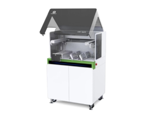
Automated Staining with the Celnovte Auto Stainer ST501
The CNT 520 Fully Automatic Special Stainer and Coverslippe takes over the tedious steps of IHC coloring. It runs set programs for Ki-67 and p16, turning out matching slides batch after batch.
With 20 slide positions and 44 reagent positions, it seamlessly accommodates diverse samples and complex protocols. Its key innovation is four independent staining stations, enabling simultaneous processing of multiple protocols without cross-contamination. This enhances laboratory flexibility and throughput. The system also supports 11 special stain protocols, meeting a wide range of diagnostic and research needs.
Automated coverslipping further streamlines laboratory operations, eliminating manual bottlenecks and boosting overall productivity. Engineered for reliability and consistency, the CNT 520 delivers high-quality staining results that empower fast-paced clinical and research environments.
Integration of Multiplex IHC for HPV Lesions
Combining Ki-67 with Other Biomarkers Like p16
Checking several markers at once paints a fuller picture of tricky tissue. In HPV-linked cervical spots, seeing Ki-67 and p16 together confirms both runaway division and broken cell-cycle guards.
Celnovte supports this integrated approach through its Multi-color immunohistochemistry, which allows dual staining on a single slide without compromising antigen integrity or signal clarity.
Benefits of Multiplexing Using Celnovte Kits
Multiplex saves precious biopsy bits and squeezes more facts from one slice. Celnovte’s kits play nicely with everyday dyes and background stains in their lineup.
Multi-color immunohistochemistry can simultaneously observe the staining results of multiple antigen targets on one slide. Through multi-color immunohistochemistry staining, the spatial relationship between different antigens is more intuitive.
Practical Considerations for Laboratory Use
Sample Handling and Antigen Retrieval Tips
To obtain clear Ki-67 signals, hidden protein sites must be gently unmasked. Using an appropriate antigen retrieval buffer can open epitopes without damaging tissue structure. Maintaining consistent heat and even temperature across all samples is also essential to ensure reproducibility during pre-treatment steps.
Quality Control in IHC Procedures
Solid daily checks keep IHC trustworthy. Always run a known busy tissue—like tonsil—alongside patient slides for Ki-67.To streamline this process, Celnovte offers a Control Slides Set, which includes pre-validated positive tissues to verify staining accuracy on a daily basis. Breast lumps can be found with internal controls, and there are special quality control products for frozen immunohistochemistry.
By combining advanced lab equipment with high-quality, customized reagents from Celnovte, laboratories can enhance their diagnostic capabilities in HPV screening. Celnovte also offers tailored solutions to meet diverse clinical needs—interested parties can contact us for more information.
FAQ
Q: What does a high Ki-67 index indicate in cervical biopsies?
A: A high Ki-67 index reflects increased cell proliferation beyond the basal layer, suggesting dysplasia or early malignant transformation often induced by HPV infection.
Q: Is Ki-67 alone sufficient for diagnosing HPV-related cervical lesions?
A: No. While helpful, using Ki-67 alone may lead to false positives. It is best used alongside p16 in dual staining protocols to improve diagnostic specificity. Celnovte’s dual-staining kits are optimized for this application.
Q: How can I optimize my lab workflow for cervical cancer screening using IHC?
A: Implementing automated platforms like the CNT 520 Fully Automatic Special Stainer along with validated antibodies like Ki-67 ensures consistent high-throughput processing with minimal variability.
Q: What tissues are recommended as positive controls for validating Ki-67 staining?
A: Tonsil or lymph node tissues are commonly used due to their high proliferative index. For ease of use, Celnovte provides ready-to-use positive control slides specific for Ki-67 detection.