
-
-
SHOP

What Every Lab Technician Should Know About Ki-67 Levels
![]() 2025-12-19
2025-12-19
By admin

Introduction: The Critical Role of Ki-67 in Pathology
Think of Ki-67 as a little flag that only pops up inside the nucleus when a cell is getting ready to divide. The higher the percentage of cells waving that flag, the faster the tissue is growing. For anyone working at the bench, this one number can change how a patient is treated, especially in oncology cases. New technicians frequently ask, “So what’s a normal Ki-67?” The real answer is always the same: it depends entirely on the type of tissue and the clinical question.
Decoding Ki-67: The Proliferation Marker
Ki-67 protein shows up the moment a cell leaves its resting phase and starts cycling through G1, S, G2, and mitosis. As soon as the cell slips back into G0, the protein vanishes. That on-off behavior makes Ki-67 a very direct snapshot of how many cells are actively proliferating right now. The only reliable way to see it is good immunohistochemistry staining, which means the quality of what happens in the lab directly affects the final score the pathologist reports.
Celnovte’s Commitment to Precision Diagnostics
Celnovte builds reagents and instruments that help pathology labs turn out sharp, repeatable results every single day. Their lineup covers IHC reagents, CISH, and FISH products that meet tough standards for sensitivity and specificity. The aim is straightforward: give labs tools they can trust so clinicians get numbers they can act on.
The Context-Dependent Nature of “Normal” Ki-67
There simply isn’t a single “normal” value that fits every case. A level that looks low in one organ can look alarmingly high in another.
Ki-67 Expression in Non-Pathological Tissue
In normal skin, you only see Ki-67 in the basal layer, where fresh cells are born. In the gut, it’s limited to the bottom of the crypts. Both places usually show fewer than 5–10% positive nuclei because that’s all the turnover a healthy body requires. When a tech runs a tonsil or appendix control and finds just a handful of dark nuclei in the right zones, everything is working as it should. Widespread staining in a “normal” control almost always means something is wrong: regeneration, inflammation, or early neoplasia.
Interpretive Variation Across Tumor Types
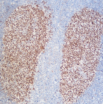
In breast cancer, Ki-67 has become a standard prognostic tool. Most labs call 0–10% low, 11–20% intermediate, and anything over 30% definitely high-grade, although the exact cut-offs still differ slightly between guidelines. A technician who gets 2% on an obviously high-grade breast carcinoma should immediately suspect under-staining, because genuine triple-negative or HER2-positive high-grade tumors almost never fall that low. The same 2% in a carcinoid or low-grade neuroendocrine tumor, however, fits perfectly. Knowing the expected ballpark for each tumor type prevents disastrous misinterpretation.
Ki-67 and p16 Dual Staining: A Case Study in Cervical Screening
One of the clearest uses of Ki-67 is in cervical cytology. When p16 and Ki-67 are stained on the same slide, any cell that lights up for both markers is almost certainly transformed by high-risk HPV. Normal squamous cells never show this combination. Celnovte released a Green/Blue p16/Ki-67 cocktail in 2023 that many cytology labs now prefer because the color contrast makes double-positive cells easier to spot than the traditional red/brown combination. No co-expression is reassuring; even one or two double-positive cells in a Pap smear usually sends the patient straight to colposcopy.

Overcoming Technical Challenges in Ki-67 Quantification
Tiny staining problems: weak antigen retrieval, foggy background, or blotchy DAB, can easily swing the counted percentage by 10–15 points. That’s enough to push a breast cancer patient from “luminal A” into “luminal B” and change her treatment plan completely.
The Need for Unsurpassed Sensitivity and Specificity
That’s why labs keep looking for cleaner detection systems that stay clean and strong. Celnovte responded in 2023 with the MicroStacker™ Plus Polymer Detection Kit.
Introducing Celnovte’s Advanced Detection Reagents
-
MicroStacker™ Plus Polymer Detection Kit: Enhanced Performance
The key is a patented compact micro-polymer design. Instead of the old bulky dextran backbone found in older polymer kits, Celnovte uses a small scaffold and stacks Fab fragments and HRP enzymes layer upon layer. The molecule ends up smaller and denser, so it penetrates crowded nuclei better and produces a sharp, dark signal even when Ki-67 expression is modest, as often happens in lymphomas. Because the system is completely biotin-free, technicians hardly ever see the nonspecific background that used to ruin breast and prostate cases. In head-to-head testing at several large reference labs in China, the MicroStacker™ Plus routinely dropped background to almost nothing while increasing the visible Ki-67 signal by 20–30% over the leading imported polymer kits.
-
P16/Ki-67 Dual Staining Solution: Streamlining Cytology
Celnovte’s p16/Ki-67 cocktails work on both ThinPrep and SurePath preparations with one 30-minute primary antibody step. The kits ship ready-to-use, and labs can choose between the classic Red/Brown and the newer Green/Blue versions. Cytology labs that switched to Celnovte’s cocktail in 2024 often report cutting their ASC-US and LSIL review rate by more than half simply because the double-positive cells stand out so clearly.
Achieving Workflow Excellence Through Automation
Staining thirty Ki-67 cases by hand day after day wears technicians out and introduces small timing or volume differences that translate into real score variation.
The Importance of Standardization and High-Throughput
Automated stainers remove almost all of that human variability and let a lab finish a large batch overnight with every slide treated identically.
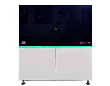
Celnovte’s CNT 360 Fully Automatic IHC&ISH Stainer
The CNT 360 runs IHC, FISH, and CISH on the same platform and can handle up to 60 slides in a single four-hour run.
-
Features: Speed and Efficiency
Techs load the trays and walk away. A full Ki-67 run from deparaffinization to counterstain takes roughly 2.5 hours for 30 slides, making same-day reporting realistic even in high-volume hospitals.
-
Advantage: Ensuring Diagnostic Consistency
Every protocol is fixed, so the staining intensity on slide 1 matches slide 60 exactly. Since 2018, Celnovte has installed over 800 of these stainers worldwide, and many customers say the rock-steady day-to-day Ki-67 reproducibility is the reason they bought additional machines.
Conclusion: Elevating Diagnostic Precision
For the technician at the bench, the message is simple: a Ki-67 percentage only means something when you know the tissue type and the reason for testing. Getting that percentage right begins with good tissue processing and ends with reagents and instruments that don’t add artifact or lose signal. Celnovte supports laboratories with exactly those tools: the MicroStacker™ Plus kit for clean, strong Ki-67 staining, p16/Ki-67 dual-stain cocktails that make cervical screening faster and more certain, and the CNT 360 stainer that turns inconsistent manual work into reliable automation. When technique and equipment both perform at their best, pathologists get scores they can trust, and patients receive the most accurate diagnosis possible.
FAQ
Q: What is the primary purpose of the Ki-67 marker?
A: Ki-67 shows which cells are actively dividing, giving pathologists a direct measure of how fast a tumor or tissue is growing.
Q: Why is consistent staining quality crucial for Ki-67 scoring?
A: The final percentage comes from counting positive nuclei by eye or digitally. Weak staining hides real proliferating cells; background creates false ones. Either error can alter patient management.
Q: What technology gives Celnovte’s MicroStacker™ Plus superior sensitivity?
A: A patented compact micro-polymer scaffold loaded with layered Fab fragments and HRP enzymes, plus a completely biotin-free design.
Q: In what clinical setting is the p16/Ki-67 dual stain most commonly used?
A: Primarily in cervical cancer screening on liquid-based Pap samples to detect transforming HPV-infected cells that routine cytology might miss.