
-
-
SHOP

Top 5 Applications of Multiplex Immunohistochemistry in Oncology
![]() 2025-05-01
2025-05-01
By admin
Multiplex Immunohistochemistry (mIHC) has become a powerful tool in cancer studies. It helps scientists and doctors examine many markers in one tissue sample. This method reveals details about tumors, boosting progress in cancer diagnosis and tailored treatments. This blog explores the top five uses of Multiplex Immunohistochemistry in oncology. It focuses on tasks like classifying tumors, studying immune cells, and finding treatment targets. These uses are shaping the future of cancer care.
Why Multiplex Immunohistochemistry Matters in Oncology
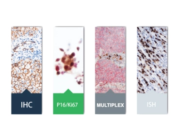
Regular immunohistochemistry (IHC) can only detect one or two markers per tissue slice. This often requires many slices, using up precious samples. However, Multiplex Immunohistochemistry detects several markers at once. It saves tissue and gives a fuller picture of cell interactions. This is vital in cancer research, where understanding tumors is key to creating effective treatments.
For researchers and doctors, mIHC offers great accuracy and works well with modern imaging and computer analysis. Below, we discuss five major ways Multiplex Immunohistochemistry is changing cancer research and medical practice.
Key Applications of Multiplex Immunohistochemistry in Oncology
Tumor Subtyping with Multiplex Immunohistochemistry
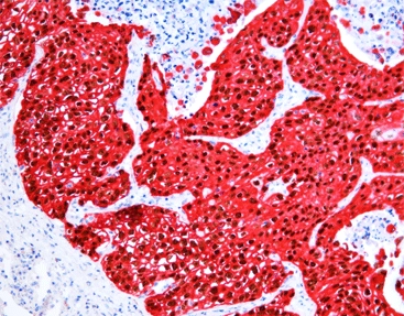
Classifying tumors is a core part of precise cancer care. It helps doctors sort cancers by their traits. Multiplex Immunohistochemistry improves this process. It detects many markers, like HER2, ER, and PR in breast cancer, in one tissue slice.
- Advantages: mIHC shows a clear view of tumor differences. It boosts diagnosis accuracy.
- Example: In breast cancer, mIHC separates types like luminal A, luminal B, HER2-enriched, and triple-negative in one slide.
- Tools: Celnovte’s Multiplex IHC Kits make tumor sorting easier with sensitive staining methods.
This method reduces the need for extra tissue slices. It saves samples and speeds up diagnosis.
Immune Cell Profiling in the Tumor Microenvironment
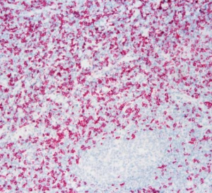
Knowing the immune setup of tumors is crucial for immunotherapy development. Multiplex Immunohistochemistry is excellent at studying immune cells, such as T-cells, B-cells, and macrophages, in the tumor area.
- Main Markers: CD3, CD8, CD20, and PD-L1 can be stained together. This shows immune cell presence.
- Effect: mIHC reveals how immune cells and tumor cells interact. It guides immunotherapy plans.
- Example: In lung cancer, mIHC spots PD-L1 patterns. This predicts how well checkpoint inhibitors work.
Celnovte’s Immune Chromogenic Reagent Double Stain I supports dual-marker staining. It improves immune cell study accuracy.
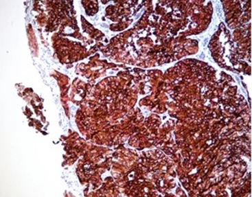
Therapeutic Target Identification
Finding workable treatment targets is a main goal in cancer research. Multiplex Immunohistochemistry helps scientists spot several druggable targets, like EGFR, ALK, and ROS1, in one test.
|
Target |
Cancer Type |
mIHC Benefit |
|
EGFR |
Lung Cancer |
Detects EGFR and PD-L1 together. This helps plan combined treatments. |
|
HER2 |
Breast Cancer |
Stains HER2 with Ki-67. It shows tumor growth rates. |
|
ALK |
Lung Cancer |
Analyzes multiple markers quickly. It confirms targets fast. |
Celnovte’s Immune Chromogenic Reagent Double Stain II provides clear staining. It ensures reliable target spotting and speeds up drug creation.
Biomarker Co-Expression Analysis
Multiplex Immunohistochemistry is perfect for studying how markers work together. This is key to understanding tumor behavior and treatment resistance. For instance, PD-L1 and IDO1 together in melanoma may show immune escape methods.
- Process: mIHC stains several markers. Then, digital imaging measures their levels.
- Result: It reveals marker patterns. This informs combined treatment plans.
- Tool: Celnovte’s Immune Chromogenic Reagent Fast Red gives bright, clear staining for marker studies.
This method helps researchers uncover complex tumor interactions.
Monitoring Treatment Response
Tracking how tumors react to therapy is vital for better patient results. Multiplex Immunohistochemistry follows changes in marker levels before and after treatment. It shows how well treatments work.
- Example: In colorectal cancer, mIHC tracks KRAS and BRAF levels. It checks targeted therapy effects.
- Benefit: One-slide testing reduces errors compared to regular IHC.
- Effect: It allows quick changes to treatmentften taken from the original. It helps doctors make smart choices for patient care.
By using mIHC, doctors can make informed decisions. This improves patient outcomes.
Celnovte: Your Partner in Multiplex Immunohistochemistry Solutions
Celnovte is a top provider of immunohistochemistry tools. It equips researchers and doctors with advanced tools for Multiplex Immunohistochemistry. Located in China, Celnovte offers high-quality antibodies, automated staining systems, and multiplex IHC kits. These ensure accuracy and consistency. Their unique MicroStacker™ technology and CNT330 Full Automatic Multiplex IHC Stainer simplify tasks. Celnovte is a trusted partner for cancer research and diagnostics worldwide. Visit Celnovte’s homepage to learn more.
Frequently Asked Questions (FAQs)
Q1. What is Multiplex Immunohistochemistry, and how does it work?
A1. Multiplex Immunohistochemistry is an advanced method. It detects many protein markers in one tissue sample. It uses specific antibodies and colored or glowing labels. The process involves applying antibodies one by one or together. Then, microscopes or digital tools show the results.
Q2. How does Multiplex Immunohistochemistry improve cancer diagnostics?
A2. Multiplex Immunohistochemistry boosts diagnostics. It gives a full view of the tumor area. This helps classify tumors, study immune cells, and analyze markers in one test. It saves tissue and improves accuracy.
Q3. What are the challenges of implementing Multiplex Immunohistochemistry?
A3. Challenges include antibody mix-ups, signal overlaps, and the need for standard methods. These can be fixed with careful antibody choices, set staining steps, and advanced imaging tools.
Q4. Why is Multiplex Immunohistochemistry preferred over traditional IHC?
A4. Multiplex Immunohistochemistry is favored because it saves tissue. It reduces errors and provides richer data. It studies many markers at once, making it ideal for complex cancer studies.
Call to Action: Elevate Your Oncology Research with Multiplex Immunohistochemistry
Are you ready to boost your cancer research with Multiplex Immunohistochemistry? Discover how mIHC can improve your work, from classifying tumors to finding treatment targets. Check out Celnovte’s product page to find advanced multiplex IHC kits and reagents suited to your needs. Contact Celnovte today to speed up your research and make breakthroughs in cancer care!

