
-
-
SHOP

The Main Challenges of IHC in Complex Tissue Analysis
![]() 2025-09-26
2025-09-26
By admin
The Intricacies of Immunohistochemistry in Complex Tissues
Complex tissues include things like tumors with mixed cell groups or well-organized organs. They create major barriers for steady and dependable IHC staining and reading.
Tissue Heterogeneity and Antigen Preservation
Complex tissues usually contain varied cell kinds, different levels of cell growth, and detailed extracellular matrices. This variety can cause uneven reagent entry and mixed antigen access. Also, the steps before analysis—like tissue fixation and handling—are key.Improper fixation can break down antigens or hide epitopes. This makes them hard to spot. On the other hand, too much fixation can link proteins tightly. This blocks antibody attachment. Keeping ideal antigen levels across various tissue types in one sample is an ongoing difficulty. It directly affects the standard and trustworthiness of IHC outcomes.
Signal Specificity and Sensitivity
Gaining high signal specificity and sensitivity in complex tissues is vital. Unwanted antibody attachment can create background interference. This hides real positive signals and leads to wrong positives. In contrast, low sensitivity means weak or rare antigens might be overlooked completely. Such antigens often appear in early illness phases or certain cell groups. This equilibrium is especially tough in tissues with strong natural enzyme action or self-glow. It needs careful tuning of methods and solid detection setups. These help separate true signals from errors.
Interpretation and Quantification
The reading of IHC results in complex tissues requires skilled assessment. Staining designs can differ a lot—nuclear, cytoplasmic, membranous, or mixed. Their strength can vary greatly even in the same section. Judging these layered staining designs demands a solid grasp of illness biology. It also calls for close focus on details.Quantifying these results objectively is another barrier. Personal judgment can bring differences between viewers. For exact diagnosis and study, the skill to correctly read and measure small changes in antigen display is key.
Innovative Solutions for Overcoming IHC Challenges
At Celnovte, we focus on creating modern items that tackle the built-in difficulties of IHC in tough tissue samples. Our full range is built to improve each part of the IHC process. This goes from choosing primary antibodies to automatic staining and modern detection.
A. Advanced Reagents for Enhanced Performance
To handle problems of specificity, sensitivity, and antigen keeping, labs need reagents that are carefully checked and tuned.
High-Quality Primary Antibodies
Our wide array of IVD-grade primary antibodies comes from both murine and human monoclonal antibody platforms. They are supported by a steady engineering expression platform. We supply a large choice, including important biomarkers like ER, PR, and HER2-PD-L1. These have steadily earned top scores in strict NordiQC reviews for several years in a row. For instance, our ER antibodies have gained Class I registration certificates cleared by NMPA. This dedication to standard guarantee sexceptional specificity and sensitivity. It cuts down background interference and provides clear, readable signals even in mixed tissue settings.
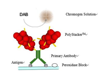
MicroStacker™ Detection Systems
Our own MicroStacker™ Detection Systems, including the Ultra Polymer Detection Kit, form a top market group in IHC. These setups use advanced polymer technology that gives notably boosted signal growth. At the same time, they lower unwanted attachment and background interference. This fresh technology helps a lot in complex tissues. There, low antigen display or strong background meddling can harm results. The build differences in our secondary antibodies for frozen sections, for example, permit greater enzyme linking. This leads to quicker and more dependable detection. This strong signal-to-noise balance makes sure that even weak positive signals show up clearly. It allows more certain and precise diagnoses.
B. Automated Staining for Reproducibility and Efficiency
Manual IHC methods are open to human mistakes and changes between groups, especially in busy settings. Our automatic tools offer an answer for consistent, reproducible, and efficient staining.
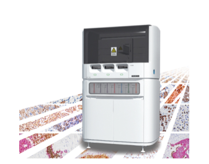
Our fully automated immunohistochemistry stainer meets the needs of modern pathology labs. With high capacity and the ability to stain 60 slides in just 2.5 hours, it significantly boosts productivity and reduces wait times. This device ensures consistent, reproducible results by precisely controlling every staining step, minimizing human error common in manual methods. It also supports new techniques like chromogenic in situ hybridization, multi-color immunohistochemistry, and p16/Ki-67 double staining, allowing labs to confidently adopt advanced diagnostics with automation.
C. Specialized Techniques for Difficult Cases
Some diagnostic obstacles need special IHC methods. These give more details or quicker outcomes than the usual ways.
Multiplex IHC (Multi-color immunohistochemistry)
For very complicated cases or limited tissue samples, our Multiplex IHC answers allow the same-time viewing of staining results for multiple antigen targets on a single slide. By showing several biomarkers at once, this method provides a more intuitive understanding of the spatial relationship between different antigens. This is key for checking tumor micro infiltration, describing immune cell groups, and boosting the overall precision of pathological diagnosis. This is especially helpful for small biopsy tissues. There, it effectively saves valuable material for full testing.
Fast Frozen IHC (Intraoperative rapid frozen immunohistochemistry)
Our Fast Frozen IHC abilities handle the pressing need for quick diagnostic info during operations. This method permits complete immunohistochemical staining operations within approximately 15 minutes. It offers instant spotting of tumor markers. This quickness is priceless for choices during surgery. It notably improves diagnostic precision for unusual shapes, tough cases, and young patients. Thus, it cuts down on delayed diagnoses. Our frozen IHC products hold special filing certificates. All of them are Class I. This makes sure that surgeons can create matching surgical plans based on timely and dependable IHC results.
Partnering for Precision Pathology
Celvote,established in 2010, is a high-tech enterprise focused on the study, creation, making, and sales of precision diagnostic instruments and reagents for tumors. Based in Zhengzhou, China, with R&D centers in Shenzhen, Suzhou, and Maryland, USA, we run GMP-compliant facilities. We are certified forISO13485, ISO9001, and possess CE-ID and NMPA approvals. We commit to supplying top-level products and services. We aim to be a standard technology-fresh enterprise in the field.
By combining our modern primary antibodies, MicroStacker™ Detection Systems, and fully automated IHC stainers, we give a full answer for handling the main obstacles of IHC in complex tissue analysis. We ask you to check our full range and find out how our fresh products can raise the precision and output of your lab work.
Conclusion
The difficulties of IHC in varied and tough tissue samples call for refined answers. By tackling issues of tissue variety, signal specificity, and reading, our modern reagents and automatic systems empower pathologists. They help achieve more precise and steady outcomes. Work with us to use cutting-edge technology. Overcome the barriers in complex tissue analysis. In the end, this adds to better patient results and scientific progress.
FAQ
Q: Are all reagents ready-to-use? Is DAB ready-to-use?
A: Most reagents are ready-to-use. DAB, however, needs to be prepared on-site, typically once every half day.
Q: How can your frozen IHC be so fast? Is there a difference in frozen reagents compared to traditional ones?
A: Our frozen IHC process involves technical adjustments and specially redeveloped reagents from fixation onwards. The breakthrough in second antibody technology, which allows for higher enzyme polymerization in frozen sections, along with reduced first antibody incubation time, contributes to its speed. The structure of these reagents differs from traditional second antibodies.
Q: Will different technicians operating the IHC stainer yield different results?
A: For technicians proficient in immunohistochemical operation steps, the staining results are consistently reproducible.