
-
-
SHOP

Inside a Lab Detection System for Accurate Results
![]() 2025-11-28
2025-11-28
By admin

The Invisible Journey: Why Precision Detection Defines Pathology
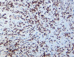
A tissue sample taken during a biopsy starts a long trip. It rides in a little jar from the operating room to the pathology lab, gets sliced paper-thin, stuck on glass slides, and finally ends up under a pathologist’s microscope. At the end of that journey, somebody has to say “cancer” or “no cancer,” and lives change based on that call. In today’s world, just looking at the shape of cells often isn’t enough. Doctors need to see specific proteins or bits of DNA that prove what’s really going on. The only way to make those invisible molecules show up bright and clear is the detection system—the part of the staining process that turns a faint whisper into a loud, unmistakable signal.
Bridging the Gap Between Sample and Diagnosis
Pathologists hate guessing. When a breast biopsy comes in and the cells look borderline, the doctor doesn’t want to send a 38-year-old mom for chemo if it’s not necessary, and definitely doesn’t want to call something “benign” when it’s not. That’s where immunohistochemistry (IHC) comes in. A primary antibody sticks to the exact protein you care about—say, HER2 or PD-L1—and then the detection system lights it up so everyone can see it. Without a strong detection step, even the best antibody in the world stays invisible. I’ve watched pathologists squint at pale, barely-there stains and shrug their shoulders. Good detection removes the shrug.
The Need for Amplification: From Molecule to Microscopy
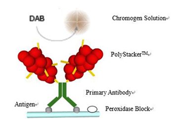
Most cancer markers hide in tiny amounts. You might have only a few hundred copies of the protein in an entire cell. The detection system’s job is to take that handful of bound antibodies and pile on dozens—or hundreds—of color-producing enzymes. When the lab adds the chromogen (basically, lab paint), those enzymes turn it into a dark, crisp spot right where the target lives. Celnovte has spent years building polymer-based systems that stack more enzymes in the right place than anybody else thought possible.
The Mechanism of Modern Detection Systems: Beyond Basic Staining
Twenty years ago, labs used clunky two- or three-step methods with biotin and streptavidin that left brown gunk all over the slide. Today, everything rides on cleaner, smarter polymer backbones.
Phase 1: Specific Target Identification (The Antibody Lock-and-Key)
It all starts with the primary antibody. Celnovte keeps more than 500 ready-to-use primaries on the shelf, including over 130 of their own cloned rabbit and mouse monoclonals. Pick the one that fits your target, drop it on the slide, and it locks on like a heat-seeking missile.
Phase 2: Signal Amplification – The Polymer Revolution
Once the primary antibody is in place, the lab adds the detection reagent. In Celnovte’s case, that’s usually something from the MicroStacker line. Picture a long, flexible polymer chain with dozens of HRP or alkaline phosphatase molecules hanging off it like Christmas lights. One end of the polymer grabs the primary antibody, and suddenly every bound antibody is carrying a whole fireworks show of enzymes. Add DAB or Fast Red and—bam—dark brown or bright red deposits pop exactly where the antigen sits. The slide goes from ghost-quiet to shouting the answer in under ten minutes. Labs that switched from old ABC kits to polymer systems typically cut background mess by half and double the signal on tough low-expressors like PD-L1 or mismatch-repair proteins.
Celnovte’s Polymer Technology: Achieving Unsurpassed Sensitivity
MicroStacker Ultra Detection Systems
The MicroStacker family has been the workhorse since the first polymer version hit labs in 2018. In 2023, Celnovte brought out MicroStacker Ultra, and most big reference labs I talk to say it’s currently the cleanest, strongest system they’ve ever run.
What makes it different in daily work:
-
It catches antigens that barely show up at all. Think nuclear Ki-67 in just a few scattered lymphoma cells or faint membranous HER2 in a breast core that looked 1+ on the old system.
-
Background stays almost snow-white even on tricky tissues like liver, kidney, or decalcified bone.
-
You can buy it as a simple two-step manual kit or in bulk bottles that drop straight into automated stainers.
I’ve had lab managers tell me they finally stopped repeating stains on “weak positives” once they moved to Ultra—saves hours a week and a lot of frustration.
Proprietary Technology: Minimizing Noise, Maximizing Signal
The trick is in how tightly Celnovte controls the size and shape of the polymer, plus a few secret blocking steps built into the reagent. Old systems sprayed signal everywhere; MicroStacker keeps it polite—only where it belongs.
Speed and Throughput: Integrating Detection with Automation
Great reagents are only half the battle. Busy hospitals need the stains done fast and exactly the same way every time.
PolyStacker Technology for Fast Frozen IHC
During surgery, the pathologist sometimes gets a frozen section, and the surgeon is standing there in scrubs asking, “Is it cancer? Are the margins clean?” Ten years ago, you could barely dream of running IHC on a frozen section—it took hours. PolyStacker cuts the whole thing to around 10–12 minutes start to finish with almost no loss in quality. Places doing Mohs surgery or intraoperative breast margins swear by it now.
CNT 360 Fully Automatic Stainer: The Workflow Engine
The CNT 360 is Celnovte’s big walk-away stainer. Load 60 slides, pick your protocols, hit start, and come back to finished racks. It runs the MicroStacker reagents perfectly every time—no tech-to-tech variation. Since 2018, Celnovte has shipped more than 800 automated stainers worldwide; the CNT 360 is the one most labs point to when they say their turnaround time dropped and their pathologists stopped complaining about inconsistent stains.
Authority and Rigor: Celnovte’s Commitment to Quality
Global Compliance and Certification Standards
Both the U.S. and China plants run under full NMPA & GMP rules. They carry ISO13485, ISO9001, FDA registration, and CE IVDR marks. That means a bottle made in Wuhan performs identically to one filled in California, and regulators on both sides of the planet have signed off.
Validating Performance: External Quality Assessment Data
Forty-seven of Celnovte’s primary antibodies have earned “optimal” or “good” scores from NordiQC—the tough independent ring trial that tests antibodies blind. When your Ki-67 or ER clone passes NordiQC year after year, pathologists know they can trust the pattern they see on the slide.
Conclusion: The Future of Diagnostics is in the Details
The detection system is the unsung hero that turns a smart antibody into a diagnosis you can bet a patient’s life on. With MicroStacker Ultra giving cleaner, stronger signals than ever, PolyStacker delivering answers while the patient is still on the table, and the CNT 360 keeping every slide perfect in high-volume labs, Celnovte has pretty much removed the weak links that used to frustrate pathologists. Better stains mean clearer calls, fewer unnecessary surgeries, and more patients getting exactly the treatment they need—no more, no less.
FAQ
Q: What is the main function of the MicroStacker Detection System?
A: It takes the tiny signal from the primary antibody and blows it up into a crisp, dark stain using a polymer loaded with enzymes—so even scarce markers show up loud and clear.
Q: How does PolyStacker Technology benefit the pathology lab workflow?
A: It knocks frozen-section IHC down to about 10 minutes with reliable staining, letting surgeons get answers before they close the patient.
Q: What evidence confirms the quality of Celnovte’s reagents?
A: Forty-seven antibodies have scored optimal or good on NordiQC external assessments, and over 800 automated stainers are running daily in labs around the world without complaints.
Q: Which automated instrument from Celnovte supports high-throughput IHC staining?
A: The CNT 360 Fully Automatic IHC Stainer handles dozens of slides at once, runs MicroStacker protocols flawlessly, and keeps every run looking identical.