
-
-
SHOP

Validating ADC Targets with IHC and Multiplex IHC (mIHC): A Practical Guide
![]() 2025-07-18
2025-07-18
By admin
Antibody–drug conjugates (ADCs) are transforming precision oncology by combining the specificity of monoclonal antibodies with the cytotoxic power of small-molecule drugs. By selectively delivering potent payloads to tumor cells, ADCs aim to maximize antitumor efficacy while minimizing systemic toxicity.
However, the clinical success of an ADC critically depends on rigorous target validation. The ideal ADC target must be highly and consistently expressed in tumor tissue while showing minimal or no expression in normal cells.
Immunohistochemistry (IHC) and multiplex immunohistochemistry (mIHC) are indispensable tools for this purpose, enabling direct visualization, localization, and quantitative assessment of target antigens within intact tissue architecture.
This guide outlines a practical workflow for ADC target validation using IHC and mIHC, highlighting how Celnovte Biotechnology solutions support accurate, reproducible, and scalable target assessment in both research and translational settings.
What Is ADC Target Validation?
ADC target validation is the process of confirming that a candidate antigen:
-
Is highly expressed in tumor cells
-
Exhibits limited or absent expression in normal tissues
-
Shows appropriate cellular localization (typically cell surface–associated)
-
Demonstrates consistent expression across patient samples
Accurate target validation ensures that an ADC can bind selectively to diseased cells, thereby improving therapeutic efficacy and reducing off-target toxicity.
IHC and mIHC are particularly well suited for this task, as they allow researchers to:
-
Visualize antigen expression directly in tissue context
-
Assess spatial distribution within tumor and microenvironment
-
Quantify expression levels at single-cell resolution
Why IHC and mIHC Matter for ADC Target Validation
Key Advantages
-
Antibody specificity verification
IHC and mIHC confirm that antibodies bind specifically to the intended antigen, reducing the risk of cross-reactivity. -
Preservation of tissue architecture
These methods reveal where the antigen is expressed within the tumor mass, stromal regions, and adjacent normal tissue. -
Multiplex immunohistochemistry benefits
mIHC enables simultaneous detection of multiple markers, allowing comprehensive evaluation of the target alongside immune, stromal, or phenotypic markers. -
Quantitative insights
Digital pathology tools transform staining intensity and distribution into quantitative metrics, supporting objective target ranking.
Together, these capabilities make IHC and mIHC foundational techniques for confident ADC target selection.
Step-by-Step Workflow for ADC Target Validation Using IHC and mIHC
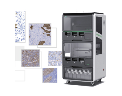
Below is a structured approach to validating ADC targets using standardized IHC and mIHC protocols, supported by Celnovte’s Multiplex Immunohistochemistry (mIHC) Kit and automated platforms such as the CNT300 and CNT330.
Step 1: Select the Target Antigen and Antibody
The first step is identifying a biologically and clinically relevant target.
-
Antigen selection
Choose antigens that are overexpressed in disease tissue (e.g., HER2 in breast cancer) and minimally expressed in healthy tissues. -
Antibody selection
Screen antibodies with high affinity and specificity. Multiple clones should be evaluated to identify the most reliable candidate. -
Pre-validation testing
Confirm antibody binding using orthogonal methods such as Western blotting or ELISA before proceeding to IHC or mIHC.
Tip:
Celnovte’s mIHC Kit allows simultaneous evaluation of 6–8 markers on a single tissue section, enabling efficient antibody specificity assessment across multiple targets.
Step 2: Prepare Tissue Samples
Proper tissue preparation is critical for high-quality staining.
-
Sample type
Use either fresh-frozen or formalin-fixed paraffin-embedded (FFPE) tissues. FFPE samples are preferred for translational and clinical studies due to long-term stability. -
Sectioning
Cut tissue sections at 4–5 µm thickness to balance morphology preservation and antigen accessibility. -
Slide mounting
Use adhesive-coated slides to prevent tissue detachment during multiplex staining.
Tissue Preparation Checklist
| Parameter | Recommendation |
|---|---|
| Tissue type | FFPE or fresh-frozen |
| Section thickness | 4–5 µm |
| Slide type | Adhesive / charged slides |
| Storage | 4 °C (short-term), −20 °C (long-term) |
Step 3: Perform IHC / mIHC Staining
This step reveals antigen expression and spatial distribution.
Typical workflow:
-
Deparaffinization and rehydration (FFPE samples)
-
Antigen retrieval using heat-induced or enzymatic methods
-
Blocking to minimize non-specific binding
-
Primary antibody incubation (single antibody for IHC, multiple antibodies for mIHC)
-
Signal detection using chromogenic or fluorescent secondary systems
-
Nuclear counterstaining (e.g., hematoxylin or DAPI)
-
Mounting for long-term signal preservation
Tip:
The CNT320 Full-Automatic Multiplex IHC Stainer automates these steps, reducing manual variability and ensuring highly reproducible results across runs.
Step 4: Image Acquisition and Data Analysis
High-quality imaging and robust analysis are essential for interpretation.
-
Imaging
Use fluorescence microscopy for mIHC or brightfield microscopy for chromogenic IHC. Whole-slide scanners enable high-throughput analysis. -
Quantification
Image analysis software can quantify antigen intensity, percentage of positive cells, and spatial relationships. -
Interpretation
Compare expression patterns between tumor and normal tissues, evaluating intensity, distribution, and cellular localization.
Example:
In breast cancer, mIHC can simultaneously visualize HER2, CD8, and PD-L1, revealing both ADC target density and immune microenvironment characteristics.
Step 5: Validation and Reporting
To ensure reliability:
-
Orthogonal confirmation using qPCR or flow cytometry
-
Reproducibility testing across multiple samples and cohorts
-
Comprehensive reporting including representative images, quantitative metrics, and interpretation criteria
Standardized protocols, such as those provided with Celnovte’s mIHC Kit, support compliance with research and clinical documentation standards.
Overcoming IHC Limitations and Maximizing Success
While powerful, IHC has inherent limitations. These strategies help optimize results:
-
Optimize antibody concentration to balance sensitivity and specificity
-
Include positive and negative control tissues in every run
-
Use automated staining platforms to minimize operator variability
-
Carefully design multiplex panels to avoid spectral overlap
-
Document all protocol parameters for reproducibility
Multiplex immunohistochemistry benefits include reduced tissue consumption, richer biological insight, and superior characterization of tumor heterogeneity compared with single-plex IHC.
About Celnovte Biotechnology
Celnovte Biotechnology specializes in the development, manufacturing, and global supply of advanced pathology diagnostics. As a leading provider of multiplex immunohistochemistry kits and automated IHC platforms, Celnovte delivers reliable solutions for both research and clinical applications.
Key products such as the CNT300 and CNT330 Full-Automatic Multiplex IHC Stainers enable high-throughput, standardized staining workflows, while the mIHC Kit supports accurate, multi-marker detection with exceptional sensitivity and reproducibility. Trusted worldwide, Celnovte’s solutions empower precise ADC target validation and translational research.

Frequently Asked Questions (FAQ)
Q1: What is ADC target validation and why is it important?
ADC target validation confirms that a target antigen is selectively expressed in diseased cells and minimally present in normal tissues, ensuring both safety and efficacy of ADC therapies.
Q2: How does mIHC differ from conventional IHC for ADC studies?
Conventional IHC evaluates one marker per slide, while mIHC enables simultaneous detection of multiple markers, providing a more comprehensive view of the tumor microenvironment.
Q3: What are common IHC limitations in ADC validation?
Limitations include single-marker analysis, potential non-specific staining, and variability in tissue processing. These can be mitigated through optimization, controls, and automation.
Q4: How can immunohistochemistry best practices improve ADC validation?
Optimized antibody titration, standardized protocols, automated staining, and rigorous controls significantly improve data reliability.
Q5: Why is multiplex immunohistochemistry preferred for ADC target validation?
mIHC provides multidimensional data from limited tissue, enabling deeper insight into target expression, heterogeneity, and tumor context.
Advance Your ADC Research with Confidence
Rigorous ADC target validation using IHC and mIHC is a cornerstone of successful antibody–drug conjugate development. By combining well-designed protocols with Celnovte’s advanced mIHC kits and automated staining platforms, researchers can generate high-quality, reproducible data that accelerates precision medicine.
Explore Celnovte’s solutions to elevate your ADC research and drive the next generation of targeted therapies.