How to Overcome TSA-Based mIHC Assay Challenges

By admin
Learning how cells and proteins work together in tissues is super important. It helps a lot in fields like cancer research and immunology. Multiplex immunohistochemistry (mIHC) is a neat tool. It lets scientists see many markers on one tissue slice at the same time. This gives a clearer picture than old staining methods. It shows tissue makeup, how cells work, how many of certain cells are there, and how cells talk to each other. This info pushes forward our understanding of tricky biological systems.
A big part of mIHC is Tyramide Signal Amplification (TSA). This method makes it easier to spot targets. It shows different markers clearly on one tissue piece. TSA works well with immunohistochemistry (IHC). It gives more details about proteins. It also makes tests more accurate and sensitive. The steps include antibodies sticking to targets, an enzyme turning on tyramide, a glowing label bonding to nearby tyrosine bits, and then removing the antibody. The glowing signal stays put. Doing this again with different glowing labels lets scientists see many targets on one slice.
Even though TSA-based mIHC is awesome, it’s not always easy to use. Scientists often face problems. Solving these issues is key to making this method work its best.
Challenges in TSA-Based mIHC Assays
TSA-based mIHC is powerful, but it comes with some tough spots. These can mess with data quality and slow down work.
Here are the main problems:
Manual Work: Old TSA methods need lots of hands-on steps. This can lead to mistakes. It also makes results vary.
Long Waits: Staining and washing many times takes a while. This can hold up research results.
Lots of Supplies: TSA supplies cost a bunch. Using them for many staining rounds adds up, especially when tweaking things.
Tricky Setup: Getting the right setup for each marker in a multiplex panel is hard. It takes time and lots of adjustments.
Fixing these problems is a must. It makes TSA-based mIHC easier to use, more reliable, and faster for research and maybe even doctor work.
Manual steps are a big hassle. But using machines can help make TSA-based mIHC smoother and fix many of these issues.
Automation: A Solution for Streamlining TSA Workflows
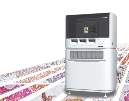
Picture a robot helper in the lab doing boring tasks. This lets scientists focus on studying results. That’s what automation does for mIHC. Machine systems tackle the problems of manual TSA steps. They cut down on hands-on time. They also make data better and more consistent. Plus, they use less supplies. Companies like Celnovte Biotech have machine stainers that make mIHC work easier and data more trustworthy.
Adding machines to TSA-based mIHC steps brings lots of good stuff. It directly fights the problems of manual ways.
Benefits of Automation
Machine systems give a steady and careful setting for the staining and cleaning cycles in TSA-based mIHC.
Here’s what’s great about them:
Clear Signals: Machines deliver supplies exactly. They also wash well. This makes signals clean with little background mess.
Even Staining: Machines stain the whole tissue slice the same way. This stops uneven spots and keeps signals steady.
Good Antibody Removal: Taking off antibodies between staining rounds is super important. It stops mix-ups and keeps results true. Machines do this well.
Keeps Tissue Nice: Machines handle tissues gently. This lowers the chance of tissue damage. It keeps the tissue shape good for study.
Same Results Every Time: Machines take out human mistakes. This makes results steady across tests and labs.
Faster Work: Machines do slow tasks like staining and washing quickly. This helps scientists get results sooner.
Less Supply Waste: Machines use just the right amount of supplies. This cuts waste and saves money, especially when setting things up.
Besides making staining better with machines, another big challenge in mIHC is removing antibodies between cycles without hurting the tissue or its ability to show markers.
Addressing Technical Challenges: Antibody Stripping
In TSA-based mIHC, you need to fully remove primary and secondary antibodies between staining rounds. This stops signals from mixing up. It also makes sure each signal is clear and correct. This cycle of adding and removing antibodies is what makes this method special. But if antibodies aren’t removed completely, signals can get messy. This ruins results.
Scientists have tried different ways to remove antibodies. Each way has good and bad parts. This shows why careful testing and tweaking are needed.
Evaluating Antibody Stripping Strategies
To remove antibodies well, you often need to break them down with heat. You also need the right pH, temperature, and solution mix. Different methods have been tested to find the best and safest way for tissues.
Common ways to remove antibodies include:
Microwave Oven-Assisted Antibody Removal (MO-AR): This uses microwave heat to break down antibodies. It works okay but can hurt delicate tissues. These tissues might peel apart.
Chemical Reagent-Based Antibody Removal (CR-AR): This uses special or custom chemical mixes to break antibodies. How well it works depends on temperature, pH, and strength. Some chemicals don’t fully remove antibodies.
Hybridization Oven-Based Antibody Removal (HO-AR): This uses a heating plate in a hybridization oven to keep a set temperature. Tests show low heat, like 50°C (HO-AR-50), doesn’t remove antibodies well. But high heat, like 98°C (HO-AR-98), works great.
Picking the best removal method is super important. This is especially true for fragile tissues where methods like microwaving can cause damage.
Recent studies have worked on gentler heat-and-chemical removal methods. These keep tissues safe while fully removing antibodies. This led to better methods like HO-AR-98.
Optimized Thermochemical Stripping Method
Knowing the limits of old methods, especially for delicate tissues, scientists made better heat-and-chemical removal processes. The Hybridization Oven-Based Antibody Removal at 98°C (HO-AR-98) is a great example. It aims to remove antibodies completely while keeping tissues in better shape than methods like MO-AR.
Using a hybridization oven at 98°C, this method fully removes primary and secondary antibodies. It works as well as MO-AR but keeps tissues, like fragile brain slices, in much better condition. This method has been used successfully in multiplex staining. It gives reliable, steady results with clear glowing signals and no mix-ups. These improved methods make TSA-based mIHC useful for more tissue types and research questions.
After staining the tissue slice well, the next big step is using smart image analysis and data handling to get useful biological info.
Challenges in Image Analysis and Data Management
Getting clear, number-based data from complex glowing multiplex images is tricky. After staining and cleaning are done, the tissue slices need to be pictured. Then, the data is processed with special computer programs.
These image analysis steps need close attention at every part, from taking pictures to getting final data. They also need checks to make sure everything is right.
Color Deconvolution and Spectral Unmixing
For mIHC and mIF, splitting signals from many markers is key for good analysis. In mIHC, color deconvolution pulls apart each colored stain from RGB pictures. For mIF, spectral unmixing is a math trick that splits glowing marker signals into separate channels. Problems include signals overlapping and tissue glowing on its own. This can make background noise. Getting spectral unmixing right is a must. It affects later steps like splitting and sorting.
Tissue and Cell Segmentation
After splitting signals, the next job is finding and marking areas in the tissue. This includes tissue parts (like tumor vs. stroma) and single cells or cell parts. This is called tissue segmentation and cell segmentation. Getting cell segmentation right is super important. It helps check where cells are, how many there are, how they interact, and where proteins are inside cells. But splitting cells is hard, especially in tissues with different cell sizes or when cells are packed tight. Mistakes in segmentation can mess up later analysis. Single-cell segmentation gives deep info, but some pixel-based methods are used too. These treat each pixel as a unit. They’re faster but don’t give cell-level details.
Phenotyping Approaches
Phenotyping means sorting cell types based on things like color, glow strength, cell shape, or a mix of these. This usually comes after cell segmentation. Sorting can use thresholding, where a cutoff value decides if a cell has a marker. Or it can use machine learning (guided or unguided). Thresholding is quick and simple but can vary between samples and users. Machine learning needs training data for guided learning or uses programs to find patterns for unguided learning. Setting clear, steady phenotyping rules is key for good results.
Making sure the programs for image analysis, like segmentation and phenotyping, are reliable is a big step in getting solid number data from mIHC/IF tests.
Algorithm Verification and Quality Control
Number results from digital pathology image tools depend on how good the programs are. So, strong quality checks (QC) for tissue segmentation, cell segmentation, and phenotyping programs are a must. Best practice rules are still being made. Checking by trained experts is super important at each step. This makes sure things work well and stay steady. Experts often review program findings by hand and fix mistakes. This helps improve the program. Number checks can also test how well programs work.
Problems still exist, especially keeping programs accurate across many slides and staining groups. These can have different staining strengths. Fixing batch differences is needed before segmentation and phenotyping. Control tissues or tissue microarrays (TMAs) stained with each batch can help make things even. Also, quality checks for whole slide images are needed. These images are often pieced together from smaller bits. Issues like image alignment, light steadiness, and lens fixes need checking. Image registration methods are also key. They align pictures from repeated staining cycles on the same slide or from nearby slices.
As the field grows, making these image analysis steps standard and ensuring data matches across labs is more important. This leads to team efforts across many groups and a need for strong data sharing.
Data Sharing and Harmonization Efforts
Sharing checked and repeatable mIHC/IF data is super important for moving research forward. This needs agreement on how data is set up and handled. Results usually include where markers are and how strong they glow for pixels or cells. Sharing raw data, final results, and main analysis code is the best way. Sharing code lets other teams redo results and try new ideas. Repositories and versioning tools make this easier. Adding sample pictures is also suggested for checking results by eye.
Team efforts across many groups are happening to make mIHC/IF steadier. Studies show good agreement between sites and within sites for some measures using the same setup and panel. These efforts aim to measure differences and set standards for reliable marker tests. This paves the way for possible doctor use. Keep standardizing across systems and learning about leftover errors are big next steps.
TSA-based mIHC/IF is a strong tool. It has changed how we study complex biological systems, like the tumor immune microenvironment. Manual staining steps have challenges, like lots of work, long times, high supply use, and tricky setups. But automation helps a lot. It makes work smoother, results steadier, and costs lower. Also, fixing technical problems like antibody removal with better heat-and-chemical methods keeps tissues safe and signals clear. Solving issues in image analysis, like segmentation, phenotyping, program checks, and batch fixes, is key for solid number data. As the field moves on, sharing data and team standardizing efforts are vital for wide use and doctor work. With more improvements in automation, method tweaks, and standardizing, TSA-based mIHC/IF will play an even bigger role in understanding diseases and making new treatments.
FAQ
Q: What are the main problems with manual TSA-based mIHC tests?
A: Manual TSA steps face issues like lots of hands-on work, which can cause mistakes and uneven results. They take a long time because of many staining and washing cycles. They use a lot of costly supplies. Also, setting up each marker right is hard and takes many tries.
Q: How does automation help fix these problems?
A: Automation cuts down on hands-on steps. This makes tests faster. It improves data quality and steadiness with even staining and good antibody removal. It also uses fewer supplies with exact dispensing. Overall, it makes work easier.
Q: Why is antibody stripping important in TSA-based mIHC, and what problems come with it?
A: Antibody stripping is key to remove antibodies from past staining rounds. This stops signal mix-ups and keeps new signals clear. Problems include removing all antibodies without hurting tissue shape or marker ability, especially in fragile tissues.
Q: What are the main steps in image analysis for mIHC/IF data, and what problems do they have?
A: Main steps are color deconvolution/spectral unmixing to split signals (problems: signal overlap, tissue glow), tissue and cell segmentation to mark areas and cells (problems: accuracy, tissue variety), and phenotyping to sort cells (problems: subjective cutoffs, training programs). Checking programs and handling batch differences are also big challenges.
Q: Why is data sharing and standardizing important in mIHC/IF?
A: Sharing data lets other scientists check results, try new ideas, and move the field forward. Standardizing steps and analysis across groups and systems ensures data matches. This improves steadiness and opens the door for possible doctor use.
RELATED PRODUCTS


