How Immunohistochemistry Unlocks Better Pathology Diagnosis

By admin
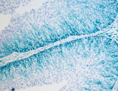
Immunohistochemistry (IHC) is a key tool in pathology labs. It helps doctors find specific proteins in tissue samples. This method makes it easier to diagnose diseases like cancer. For medical experts, using IHC panels for different body parts is important. It improves patient care.
img.How Immunohistochemistry Unlocks Better Pathology Diagnosis.webp
This blog explores common IHC panels for various organs. It shows how they boost diagnostic accuracy. We also introduce Celnovte Biotech, a leading name in multiplex immunohistochemistry (mIHC) solutions. Plus, we answer common questions to help you understand this vital diagnostic tool better.
Understanding Immunohistochemistry Basics
IHC uses antibodies to spot proteins in tissue slices. This helps identify cancers, inflammation, and other conditions. It shows how diseases work. Pathologists can tell if a growth is harmless or harmful. They can also find where a tumor comes from and check its growth markers. This leads to better treatment plans.
For doctors needing reliable tools, Celnovte Biotech offers advanced products. Their HRP Blue Stain Reagent Kit and HRP Green Stain Reagent Kit show multiple proteins in one sample. These tools save time. They also give clear results. They are essential in today’s busy pathology labs.
IHC Panels for Different Body Systems
Here, we list IHC panels used to diagnose diseases in various body parts. These panels target specific proteins. They make disease identification clear and accurate.
Head and Neck
Cancers in the head and neck, like squamous cell carcinoma or nasopharyngeal cancer, need precise IHC panels. Below are key panels for specific conditions:
- Retinoblastoma. A 12-protein panel includes CK, Vim, S-100, GFAP, CD56, Syn, SMA, LCA, p53, RB, MDM2, and Ki-67. It gives a full view of the tumor.
- Meibomian Gland Cancer. Uses 11 markers, such as BerEP4, p63, CK7, CK5/6, CK8/18, EMA, CEA, S-100, p53, BCL2, and Ki-67. These help separate it from other tumors.
- Nasopharyngeal Cancer. An 8-marker panel (CKpan, CK5/6, p63, LCA, EGFR, Ki-67, p53) with EBER testing confirms the diagnosis.
- Carotid Body Tumor. A 10-marker set (Syn, CgA, S-100, CK, EMA, Calcitonin, TTF-1, HMB45, RCC, Ki-67) spots this rare growth.
These panels work better with Celnovte’s mIHC staining tools. They let pathologists see many proteins at once. This speeds up diagnosis.
Salivary Gland
Salivary gland tumors, like mucoepidermoid or adenoid cystic cancers, need special IHC panels. These tell apart gland and muscle-like cells:
- Mucoepidermoid Cancer. Combines gland markers (CKpan, CK7, CK18, CD117, AAT, AACT) with muscle-like markers (p63, CK5/6, S-100) and Ki-67. Special stains like AB and PASD help too.
- Adenoid Cystic Cancer. Uses gland markers (CKpan, CD117, EMA), muscle-like markers (p63, SMA, S-100), MYB, and Ki-67. Extra tests may be needed.
- Acinic Cell Cancer. Includes CK7, AAT, AACT, SOX10, S-100, CD117, DOG1, p63, Calponin, and Ki-67. PASD staining confirms the diagnosis.
Celnovte’s mIHC stainer makes applying these panels simple. It supports fast, accurate results in busy labs.
Lung, Pleura, and Mediastinum
Lung and chest tumors, like lung adenocarcinoma or thymoma, rely on targeted IHC panels:
- Lung Adenocarcinoma. An 8-protein panel (CEA, CD15, CD10, CyclinD1, p53, CD34, IV collagen, Ki-67) finds early-stage lesions.
- Neuroendocrine Cancer. Uses CD56, CgA, Syn, TTF-1, CK, and Ki-67 to confirm nerve-like features.
- Malignant Mesothelioma. Needs two positive markers (Calretinin, WT1, D2-40, CK5/6, MC) and two negative ones (TTF-1, PAX8, CEA, CD15, CD117).
These panels work well with Celnovte’s reliable staining tools. They ensure strong results in tough cases.
Digestive System
The digestive system covers many cancers, from esophageal to colorectal:
- Esophageal Squamous Cancer. Uses CK5/6, p63/p40, CK7, CK19, CK20, CEA, CD56, CgA, Syn, p16, p53, and Ki-67.
- Gastric Cancer. Includes CK7, CK20, CEA, CDX2, E-Cadherin, gut markers (SATB2, MUC2, CDX2, CD10), stomach markers (MUC1, MUC5AC, MUC6), HER2, p53, and Ki-67. Extra tests (EBER, MSI, E-Cad, p53) may help.
- Liver Cancer (HCC). Diagnosed with GPC3, HSP70, and GS. Two positive markers confirm HCC.
Celnovte’s HRP Blue Stain Reagent Kit makes these markers clear. It helps tell tumor types apart.
Urological System
Urological tumors, like kidney or bladder cancer, need specific panels:
- Kidney Clear Cell Cancer. Uses CAIX, CK7, AMACR, CD117, TFE3/TFEB, HMB45, and MelanA. FISH testing may confirm.
- Urothelial Cancer. Includes GATA3, p63, 34βE12, CK7, CK20, CD44, β-catenin, p53, and Ki-67. Extra markers (CDX2, HCG, CD138) help with specific types.
- Bladder Adenocarcinoma. Uses GATA3, p63, 34βE12, CK7, CK20, CDX2, β-catenin, PAX2/PAX8, PSA, and Ki-67.
Male Reproductive System
Prostate and testicular tumors depend on IHC for clear classification:
- Prostate Cancer. Separates benign from malignant with p63, 34βE12, and AMACR.
- Testicular Seminoma. Uses SALL4, OCT4, CD117, D2-40, SOX2, CD30, LCA. FISH testing may confirm.
Female Reproductive System
Female cancers, like cervical or ovarian, need thorough IHC panels:
- Cervical Squamous Cancer. Uses p40, CK5/6, p16, p53, Ki-67, and HR-HPV testing.
- Ovarian Serous Cancer. Includes PAX8, WT1, p53, NapsinA, ER/PR, Vim, CK7, CK20, and more.
- Endometrial Cancer. Uses PAX8, p53, p16, ER, PR, β-catenin, AMACR, NapsinA, HNF1β, WT1, and MMR testing.
Celnovte’s mIHC solutions support these complex panels. They allow multiple markers to be seen at once. This ensures exact diagnosis.
Celnovte Biotech: Advancing Pathology Tools
Celnovte Biotech focuses on creating and sharing advanced pathology tools. As a leader in mIHC, they offer products like the HRP Green Stain Reagent Kit and automated mIHC stainers. These tools improve accuracy. They also speed up lab work. Celnovte is a trusted name for pathologists aiming for top results.
Common Questions Answered
Q1: Why is Ki-67 used so often in IHC panels?
A: Ki-67 shows how fast tumor cells grow. It helps doctors understand how aggressive a cancer is. It also gives clues about the patient’s outlook across many cancer types.
Q2: How does mIHC make diagnosis faster?
A: mIHC spots multiple proteins in one tissue sample at once. This saves time. It also makes results more accurate than single-protein tests.
Q3: What proteins are key for lung adenocarcinoma diagnosis?
A: Key proteins include CEA, CD15, CD10, CyclinD1, p53, CD34, IV collagen, and Ki-67. They help find early lesions. They also guide treatment plans.
Boost Your Diagnostic Skills
For pathologists and doctors, mastering IHC is crucial. It leads to better diagnoses and patient care. Using advanced IHC panels and tools from Celnovte Biotech can improve your lab’s work. Explore Celnovte’s mIHC products today. Transform your diagnostic process. Achieve top accuracy in your results.
RELATED PRODUCTS


