
-
-
SHOP

Beyond Conventional Polymers: How MicroStacker™ Technology is Redefining IHC Sensitivity
![]() 2026-03-12
2026-03-12
By admin
In today’s pathology labs, the line between a correct diagnosis and an overlooked finding can come down to how clearly one slide shows up under the microscope. As precision medicine moves forward, pathologists need sharper and more reliable results from Immunohistochemistry (IHC) than ever before. Still, many labs continue to wrestle with the built-in drawbacks of older detection methods.
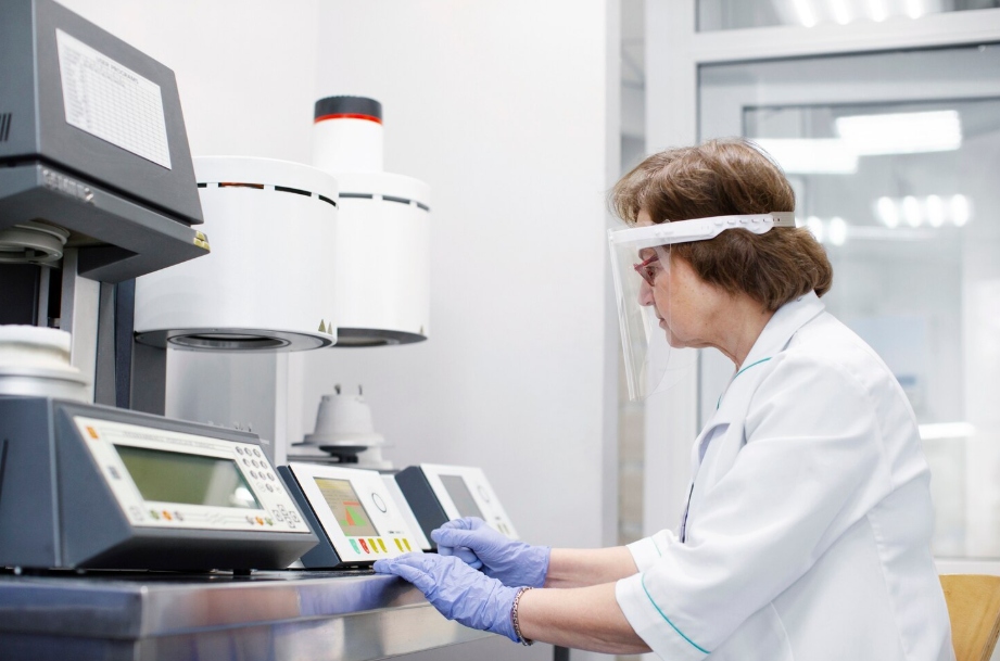
Celnovte, a company with more than 30 years of experience working in pathological diagnostic reagents and instruments, is shifting that picture with its own MicroStacker™ technology. Instead of depending on the heavy dextran carriers that have been standard for a long time, MicroStacker™ brings a fresh approach that improves signal clarity and builds greater trust in diagnostic reads.
The Challenge of Precision: Why Traditional IHC Polymers Fall Short
For a long time, the main way to detect signals in IHC has involved horseradish peroxidase (HRP) polymers anchored on big, sprawling dextran chains. Those systems marked real progress over earlier techniques, yet they also brought along several stubborn problems that current labs still have to face.
The Steric Hindrance of Bulky Dextran Backbones
Older polymers act like oversized “molecular bushes,” with antibodies and enzymes hanging off a thick, branched dextran backbone. Their sheer size creates steric hindrance. The large structure simply cannot squeeze into narrow tissue spaces or reach weakly expressed antigens. Weak signals follow, and in important cases, that can mean false negatives.
The Noise Problem: Non-specific Staining and Endogenous Interference
Big polymers tend to get trapped inside the tissue structure, which causes extra background staining, often called “noise.” On top of that, many classic systems use complete IgG antibodies that stick non-specifically to endogenous Fc receptors present in the tissue. In organs like the liver or kidney, where endogenous biotin levels run high, traditional setups also pick up unwanted staining. Pathologists then struggle to tell real positive marks from background clutter.
Introducing MicroStacker™: The Engineering of a Compact Scaffold
Celnovte tackled these long-standing issues by creating MicroStacker™ technology, which relies on autonomous kernel methods to build a smarter and more efficient molecular design.
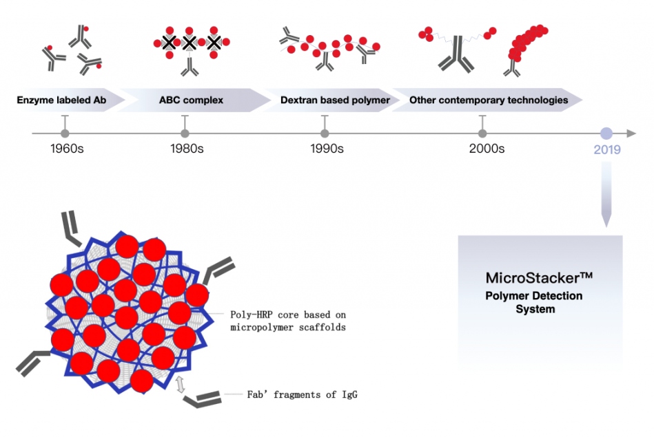
The Science of Layered F(ab’) Stacking
Rather than the scattered branching seen in dextran, MicroStacker™ uses a carefully managed, layered arrangement of F(ab’) fragments from IgG secondary antibodies and peroxidase enzymes attached to a small polymer base. This produces a tight, compact structure that holds far more signaling units per size compared with standard HRP polymers.
Maximizing Signal-to-Noise Ratio for Crystal Clear Results
Because MicroStacker™ polymers stay small, they slip more easily through tissue and reach antigens located in the nucleus, cytoplasm, or on cell membranes—places where bulky polymers often fail. Switching to F(ab’) fragments instead of whole IgG cuts out unwanted binding to Fc receptors. Removing biotin from the system also wipes away any chance of background staining from endogenous biotin. Together, these changes deliver a much stronger signal-to-noise ratio, which translates into sharper, steadier, and more vivid staining for both research and clinical use.
Celnovte’s MicroStacker™ Portfolio: Tailored Solutions for Complex Diagnostics
Celnovte has turned this core advance into a range of targeted products built to handle the tough requirements found in clinical and research settings.
MicroStacker™ Plus Polymer Detection Kit: Reaching the Pinnacle of Detection Sensitivity
The MicroStacker™ Plus HRP-Polymer Detection Kit offers a biotin-free option made for detecting mouse and rabbit primary antibodies on FFPE tissue sections. By increasing the density of enzymes and antibodies layered onto the micro-scaffold, the kit reaches higher sensitivity levels. Labs find it especially useful when confirming antigens such as HER2, TROP2, or c-Met, which matter greatly for selecting patients who might benefit from targeted treatments.
MicroStacker™ Mouse-on-Mouse Polymer Detection Kit: Solving the Endogenous Background
Staining mouse tissue with mouse monoclonal primary antibodies has always presented a major hurdle. Normal secondary antibodies cross-react with the mouse antibodies already present in the tissue, producing a heavy background. The MicroStacker™ Mouse-on-Mouse kit includes a specially designed anti-mouse secondary that blocks this cross-reactivity. That makes the kit valuable in cancer studies, particularly when researchers need clean, accurate biomarker mapping in mouse xenograft models.
Bridging Technology and Clinical Practice: Real-World Applications
The real strength of MicroStacker™ shows up in everyday lab and clinical work, where it directly tackles common workflow and interpretation difficulties.
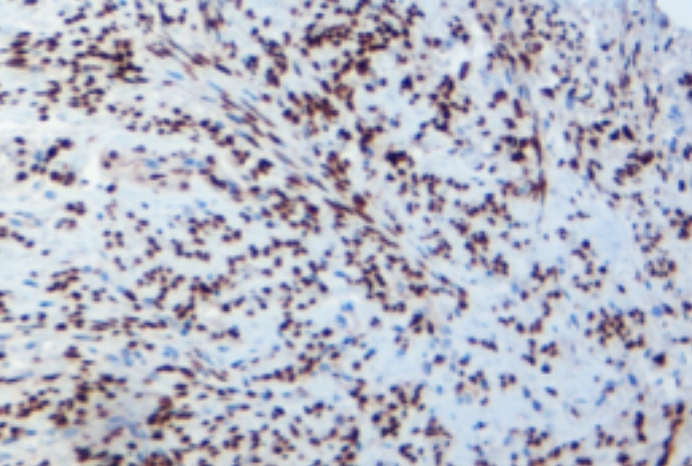
Enhancing Multiplex IHC (mIHC) Performance
Studying the tumor micro-environment often requires looking at several biomarkers together on one slide. Celnovte’s mIHC solutions, built around MicroStacker™, support simultaneous detection of multiple markers while using less tissue. Side-by-side comparisons have shown that Celnovte staining frequently appears stronger and shows clearer cellular details than results from platforms such as the Akoya Opal system. That level of detail helps pathologists better understand how immune cells and tumor cells sit next to each other.
Optimizing Low-Expression Antigen Detection in Rare Tumors
Rare tumors or cases with unclear cell shapes demand detection that picks up even very faint protein signals. The high sensitivity of MicroStacker™ makes sure those subtle expressions do not get missed. For example, key studies have used Celnovte antibodies to detect a new COX7C::ALK rearrangement in lung tumors, highlighting the need for dependable sensitivity to reach the right diagnosis. In fast-paced intraoperative settings, the same high-sensitivity detection can deliver reliable staining in just 10-15 minutes, giving surgeons the information needed to decide on the spot.

Proven Authority: Data-Driven Reliability
Celnovte backs its claims with solid outside testing rather than simple statements. The company offers more than 460 primary antibodies, and a good number have received optimal or good marks in NordiQC evaluations. In particular, self-developed antibodies for ER, PR, and HER2 have held “optimal” scores for six years running, which reflects the steady performance of both the antibodies and the paired detection systems.
Celnovte maintains its main office in Maryland, USA, and runs GMP-compliant production sites in China. The company holds ISO13485, ISO9001, and CE IVDR certifications, so every product meets strict international quality standards.
Transform Your Lab with MicroStacker™ Technology
Do you want to cut down on background staining and gain better sensitivity in your IHC work? Consider the advantages of a smaller, more efficient polymer design. Reach out to Celnovte now to arrange a demonstration or obtain a product brochure. See for yourself how MicroStacker™ can sharpen diagnostic accuracy in your laboratory.
FAQ
Q: Why is the signal-to-noise ratio more important than absolute signal strength in IHC?
A: Strong staining loses value if background noise drowns it out. A high signal-to-noise ratio makes specific staining stand out clearly from artifacts, which improves both digital quantification and manual reading by pathologists.
Q: How does the removal of the biotin-streptavidin system improve lab efficiency?
A: Labs no longer need a separate step to block endogenous biotin. That shortens the overall protocol, lowers reagent use, and prevents false background in tissues such as liver or kidney that naturally contain high biotin levels.
Q: Is MicroStacker™ technology compatible with automated staining platforms?
A: Yes. MicroStacker™ works smoothly on automated IHC stainers, including the CNT 360, delivering consistent, high-volume, and repeatable results suited to demanding clinical labs.
Q: Can compact polymer technology improve the detection of nuclear targets?
A: Yes, it can. The reduced steric hindrance of compact polymers like MicroStacker™ Plus allows better access to nuclear sites, even when antigens sit in crowded or small spaces. That leads to reliable staining across nuclear, cytoplasmic, and membranous locations.
![[Instrument] MicroStacker™ Plus Polymer Detection Kit](https://www.celnovte.com/wp-content/uploads/2020/08/3.gif "[Instrument] MicroStacker™ Plus Polymer Detection Kit")
![[RX] MicroStacker™ Mouse-on-Mouse HRP Polymer](https://www.celnovte.com/wp-content/uploads/2021/05/1.gif "[RX] MicroStacker™ Mouse-on-Mouse HRP Polymer")