
-
-
SHOP

Lung Cancer Biomarkers & IHC Testing: A Pathological Diagnosis Guide
![]() 2025-10-18
2025-10-18
By admin

Nowadays, with the advent of personalized medicine, the detection of lung cancer involves more than just tissue typing. In the current era, understanding the tumor’s individual molecular features is crucial in determining specific treatment and predicting patient outcomes. For oncologists and pathologists, it implies the identification of certain markers in tumor specimens. Immunohistochemistry (IHC) is a key method in this case, offering a strong, visual tool to reveal the important information contained in each cell. This overview will cover the major lung cancer markers and illustrate how a good, reliable IHC process is at the core of obtaining an accurate diagnosis.
Understanding Lung Cancer Biomarkers
What Are Biomarkers?
A biomarker is a definitive sign of a biological state or condition. Cancer biomarkers are unique proteins or DNA mutations found in cancer cells. Identifying these markers enables precise tumor classification, prediction of tumor behavior, and, most importantly, determination of whether a patient could benefit from targeted therapies or immunotherapy.
NSCLC accounts for most lung cancer cases, and treatment strategies have evolved rapidly with marker-driven therapies. While many markers exist, several primary markers are routinely evaluated in pathological examinations.
• PD-L1 (Programmed Death-Ligand 1): It assists in the evasion of the immune system by cancer cells. Elevated levels of PD-L1 expression, as found by IHC, could mark a patient’s likely good response to immune therapy (immune checkpoint inhibitors).
• ALK (Anaplastic Lymphoma Kinase): ALK gene mutations form an abnormal combined protein that promotes cancer cell proliferation. IHC is a screening tool to detect the levels of ALK protein, which identifies the patients who would be candidates for ALK blockers.
• EGFR (Epidermal Growth Factor Receptor): EGFR gene mutations are frequent causes in certain forms of NSCLC. While gene testing is the ideal way to detect changes, IHC may assist in noting the presence of the protein.
• ROS1: Much like ALK, shifts in the ROS1 gene form a combined protein that boosts cancer. IHC provides a quick and useful initial tool for ROS1-positive growths.
• TTF-1 (Thyroid Transcription Factor-1) & p40: These serve as type indicators. TTF-1 usually shows up in adenocarcinoma, whereas p40 marks squamous cell carcinoma specifically. Applying an IHC set with these indicators is essential for correctly typing NSCLC.
The Role of Immunohistochemistry (IHC) in Lung Cancer Diagnosis
How IHC Works: Visualizing Proteins in Tissue
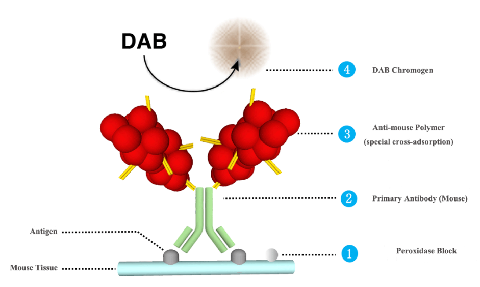
IHC serves as a lab method that employs antibodies to precisely attach to aims in tissue specimens. For cases of lung cancer, this approach lets pathologists see the existence, spot, and amount of particular indicators within a growth. A primary antibody straightaway connects to the target protein. Next, a secondary antibody joins to an enzyme. This one then links to the primary antibody. As soon as a substrate gets introduced, the enzyme creates a tinted deposit right at the indicator’s place. You can view this under a microscope.
The Critical Steps of the IHC Workflow
A precise IHC outcome hinges on excellence at each stage, from handling tissue to final review. Any single flaw can weaken the whole diagnosis.
1. Tissue Preparation and Sectioning: The method starts with a formalin-fixed, paraffin-embedded (FFPE) tissue block. A top-grade microtome cuts very thin slices (often 4-5 micrometers). The standard of this first slice is crucial, since a flawed cut with rips or uneven depth can cause staining flaws and wrong readings.
2. Staining and Detection: The tissue slide next goes through phases like removing paraffin, adding water, and retrieving antigens before the staining steps start. This is where the grade of your supplies and the steadiness of your equipment matter most.
3. Analysis: A pathologist looks at the colored slide with a microscope or through a digital view to rate the marker’s presence. This gives the doctor useful diagnostic details.
Optimizing Your Lab’s IHC Workflow with Celnovte Solutions
At Celnovte, we recognize that trust in diagnosis stems from a base of quality and steadiness. We supply a full array of items crafted to help pathology labs produce accurate and repeatable outcomes.
High-Performance Reagents for Unmistakable Results
The sharpness of an IHC color ties directly to the standard of the antibodies and spotting systems in use.
• Primary Antibodies: Our collection features a broad selection of clinically important Primary Antibodies for cancer identification. Each one undergoes strict checks to confirm a strong focus and bond to its aim, cutting down stray signals and yielding sharp, distinct coloring that is simple to assess.
• MICROSTACKER™ Detection Systems: To boost the signal from the main antibody, a solid spotting system is necessary. Celnovte’s MICROSTACKER™ Detection Systems are built for excellent awareness and focus. This approach guarantees that even faintly present markers show up clearly, yielding sturdy results you can rely on and avoiding missed positives. We also provide a complete set of Ancillaries to aid each part of the coloring process.
Automation for Consistency and Efficiency
Doing stains by hand risks mistakes from people and differences, which can affect diagnostic correctness. Making this automatic is vital for current pathology.
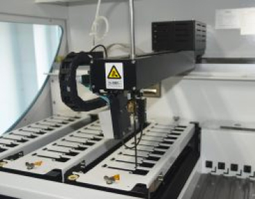
Automated IHC Stainer: The Celnovte IHC Stainer aims to simplify your lab’s operations, boost output, and secure total uniformity from slide to slide and batch to batch. By handling the full coloring process automatically, our device cuts manual effort, reduces supply loss, and removes the unevenness common in hand methods. This results in quicker delivery for urgent diagnoses and lets your trained staff tackle tougher jobs.
A Comprehensive Partner in Pathology
Celnovte goes beyond being just a supply source. We deliver an all-around option for the up-to-date pathology lab. Our item range covers tools and supplies for the whole histology flow, from basic H&E Stainers and Special Stainers to advanced choices like Multiplex IHC and Digital Scanners for digital pathology. By blending Celnovte options, you form a smooth, productive, and trustworthy diagnostic route.
Conclusion: Empowering Precision Diagnosis in Lung Cancer
Exact marker checking with IHC is vital in today’s handling of lung cancer. It enables doctors to choose informed care paths, offering patients the strongest shot at good results. Reaching this calls for a mix of pathologist skill and a top-performing, fully tuned lab process. By teaming with Celnovte, you obtain access to better supplies, steady automation, and a full backing setup focused on your success.
Ready to boost your lab’s IHC skills? Check our clinical options or reach out to our group today to discover how Celnovte can assist you in reaching diagnostic superiority.
FAQ
Q: Why is high-quality IHC so critical for lung cancer?
A: Accurate IHC testing directly impacts patient treatment. It identifies which patients will benefit from specific, life-saving targeted therapies and immunotherapies, forming the cornerstone of personalized cancer care.
Q: What makes Celnovte’s IHC solutions reliable?
A: Our reliability comes from a multi-faceted approach: high-specificity primary antibodies, ultra-sensitive MICROSTACKER™ detection systems that ensure clear signals, and automated IHC stainers that provide unmatched consistency and eliminate manual variability.
Q: Do you offer technical support for your products?
A: Yes, we provide comprehensive technical support to ensure your lab can seamlessly integrate and optimize our solutions for the best possible performance and results.