
-
-
SHOP

Understanding the Function of CISH in Pathology
![]() 2025-12-18
2025-12-18
By admin

Pathology today sits right where old-school cell shapes meet brand-new gene information. Doctors still need to look at how cells appear under the microscope, but more and more, they also need to know exactly what is going on inside those same cells at the DNA or RNA level. That combination decides whether a patient gets a targeted drug or not. Chromogenic In Situ Hybridization (CISH) is the tool that ties those two worlds together. It puts a colored dot exactly where a certain piece of DNA or RNA lives, and it does that without wrecking the picture of the tissue. You end up with a regular glass slide that shows both the molecular answer and the usual cell details side by side.
Celnovte started in 2010 and has spent every year since building reagents and machines for IHC, CISH, and FISH. The whole point is to give labs products they can count on when cancer diagnoses have to be dead-on accurate.
The Imperative for Precision: Locating Molecular Targets in Pathology
The Diagnostic Gap: Integrating Morphology with Genomics
Most cases still begin with a nice H&E slide and a handful of IHC stains. Those are great for figuring out what kind of tumor you are dealing with. Yet when the oncologist asks, “Will this breast cancer respond to trastuzumab?” or “Is this lymphoma driven by EBV?” the protein stains alone usually can’t answer. You have to prove the gene is really amplified or the virus is really there, and you have to prove it only in the bad cells, not in the normal ones mixed in. That’s the gap CISH fills.
Introducing Chromogenic In Situ Hybridization (CISH)
CISH takes a small piece of labeled DNA or RNA (the probe), lets it stick to its matching sequence on the slide, and then turns that spot into a visible dark dot with an enzyme and a chromogen. The dot stays exactly where the target is. Because everything happens on a standard paraffin section, the pathologist can look at the dots and the cell shapes at the same time under a regular light microscope.
Defining the Core Function and Operational Advantages of CISH
Bridging the Gap: Visualizing Nucleic Acids In Situ
Picture a breast cancer biopsy with scattered HER2 amplification. Some cells have twenty copies, others have the normal two. CISH puts a cluster of black dots only in the nuclei that actually carry the extra copies. The pathologist can draw the tumor border on the H&E, flip to the CISH slide, and know instantly which parts of the tumor are truly HER2-positive. That one-to-one match between gene status and cell type is the main job CISH does every day.
Functional Benefits: Brightfield Readout and Permanent Archiving
The biggest day-to-day win is that nobody needs a fluorescence microscope. Labs already have plenty of brightfield scopes, and technicians are comfortable with them. The colored dots do not fade. You can pull a CISH slide out of the file ten years later, and it still looks exactly the same. That permanence makes consults, tumor boards, and quality checks a lot easier than with FISH slides that go dark after a few months.
Celnovte’s High-Performance Molecular Reagents for CISH
Celnovte keeps a full shelf of CISH and FISH probes, plus the newer SuperISH™ line for tougher RNA jobs.
Elevating Sensitivity: SuperISH™ RNA In-Situ Hybridization Technology
DNA targets such as HER2 or ALK are usually present in hundreds or thousands of copies, so regular CISH sees them easily. RNA targets, especially viral transcripts or low-copy mRNAs, can be much scarcer. SuperISH™ is Celnovte’s home-grown answer that came out in 2020. It pushes sensitivity high enough to show single RNA molecules as individual dots, all while keeping the same brightfield readout labs already use.
Clinical Specificity: The Kappa/Lambda ISH Probe for Lymphoma Diagnostics
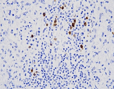
When a lymph node is packed with atypical lymphoid cells, the question is almost always “reactive or lymphoma?” Normal B-cells make both kappa and lambda light chains; lymphoma cells usually make only one kind. Celnovte’s Kappa/Lambda Probe Kit (ISH) stains kappa in one color and lambda in another on the same slide. If almost every cell lights up for only one color, clonality is obvious. The fact that the signals sit right on the nuclei of the weird-looking cells removes any doubt.
Optimizing the CISH Workflow with Digital Solutions
The Need for High-Fidelity Scanning in Molecular Pathology
Counting dots in fifty or a hundred nuclei by eye takes time, and different pathologists sometimes come up with slightly different scores. Once the slide is scanned, the software can do the counting in seconds, and everybody gets the same number.
CNT 160 Digital Slide Scanner: Precision in CISH Image Capture
Celnovte launched the CNT 160 in 2020 for exactly this job. It grabs brightfield images at high resolution, handles the strong contrast of chromogenic dots without blooming, and feeds the files straight into whatever viewing or analysis platform the lab uses. Remote sign-out becomes simple, second opinions happen with a click, and the scanned images never fade or break, as glass slides can.

Quality and Authority: Celnovte’s Commitment to Diagnostic Excellence
Global Compliance and Certifications
Plants in China and the USA run under NMPA & GMP rules and carry ISO13485 and ISO9001 certificates. The company also holds FDA registration and CE IVDR marking, so the same box of probes can ship to any major market without extra paperwork.
Validation Through Scientific Rigor and Global Reach
Forty-seven of Celnovte’s IHC antibodies have scored optimal or good on NordiQC runs; the same research and cloning teams that built those antibodies also develop the CISH and SuperISH™ probes. That track record matters when a lab has to defend a HER2 CISH result in court or during an accreditation visit. Today, our company’s reagents and machines are in routine use in more than 2300 leading hospitals across China and in over 40 countries worldwide. Labs keep coming back because the dots are where they are supposed to be, day after day.
Conclusion: CISH as the Visual Standard for Personalized Medicine
CISH turns raw gene information into something a pathologist can actually see in the exact cells that matter. It costs less than FISH, works on the scopes labs already own, and produces slides that never fade. With sharper reagents like SuperISH™ for tricky RNA targets and solid digital backup from scanners such as the CNT 160, Celnovte keeps giving labs the tools they need to turn molecular results into clear yes-or-no answers that guide treatment. That is why CISH has become the everyday workhorse for gene amplification studies, viral detection, and clonality testing, and many emerging single-cell RNA assays in hospitals around the world.
FAQ
Q: What is the primary function of CISH in pathology?
A: CISH puts a permanent colored dot on specific DNA or RNA sequences right inside the tissue, so the pathologist can match molecular findings to the cell shapes on the same slide.
Q: What type of microscope is required to read CISH results?
A: Just a regular brightfield microscope; no dark room or special filters needed.
Q: Which Celnovte product helps standardize the CISH interpretation workflow?
A: The CNT 160 Digital Slide Scanner turns glass CISH slides into permanent digital files for easy counting, storage, and remote review.
Q: How is CISH different from FISH?
A: CISH uses chromogenic (colored) dots instead of fluorescence. The slides can be viewed on any brightfield microscope, do not fade over time, and can be archived permanently—making them easier for routine diagnostics, consults, and long-term storage.