
-
-
SHOP
 PRODUCT CATEGORY
CONTACT US
![]() Phone
Phone
![]() Email
Email
![]() Address
Address
6 Cuizhu St, Zhong Yuan Qu, Zheng Zhou Shi, He Nan Sheng, China, 450001
![[Red/Brown] P16/Ki-67 Dual Staining Detection Kit](https://www.celnovte.com/wp-content/uploads/2020/08/2-8.gif "[Red/Brown] P16/Ki-67 Dual Staining Detection Kit")
![[Red/Brown] P16/Ki-67 Dual Staining Detection Kit](https://www.celnovte.com/wp-content/uploads/2020/08/3-6.gif "[Red/Brown] P16/Ki-67 Dual Staining Detection Kit")
[Red/Brown] P16/Ki-67 Dual Staining Detection Kit
P16/Ki-67 dual staining detection reagent kit (immunocytochemistry) is an detection reagent for cervical cell screening shunt and diagnosis of cervical tissue samples. It adopts immunohistochemical double-staining detection and multi-antibody combination technology. Two antigen targets, p16 and Ki-97, can be detected simultaneously on the same sample tissue section by one staining process.
The p16 gene has the function of inhibiting cell proliferation during the cell cycle arrest period and is strongly expressed in cervical intraepithelial neoplasia and cervical cancer, which is an objective biomarker for cervical lesions. Ki-67 is a biomarker of cell proliferation, and its overexpression indicates that the cell is in the proliferation phase. Normally, the expressions of p16 and Ki-67 are mutually exclusive. The co-expression of p16 and Ki-67 indicates that the cell cycle is disordered and the cells continue to proliferate. p16 alone as the identification standard is required combining morphology and detected simultaneously with Ki67. The results are more reliable.
Product Features
Application
– Combined screening with Liquid-based cytology + HPV + immunohistochemistry double staining
– As a shunt detection measure for ASC-US and LSIL
– For shunting samples with normal cytology/positive HPV test
– Shunt high-risk HPV-positive patients except HPV16/18 on HPV primary screen
Criteria for Kit Identification
Positive refers to observing the same target sample under a microscope, which can be seen single or multiple cells of the cytoplasm stained brownish yellow and the nucleus of the same cell stained red or reddish brown, that is, the same cell p16 and Ki67 are expressed simultaneously.
Negative refers to observing the same target sample under a microscope, which can not be seen single or multiple cells of the cytoplasm stained brownish yellow and the nucleus of the same cell stained red or reddish brown, that is, the same cell has only p16 expression or only Ki67 expression, or two indicators.
Specification
|
Model Number |
Name |
Specification |
Content |
|
SD9001 |
P16/Ki-67 Dual Staining Detection Kit (Immunocytochemistry) |
10mL; 30mL |
Reagent A: Peroxidase Blocking Reagent B: Combined primary antibody Reagent C: Combined secondary antibody Reagent D1: DAB substrate Reagent D2:substrate buffer Reagent E1: Fast Red Chromogenic Reagent Reagent E2: Activator Reagent E3: Fast Red Buffer Reagent F: Hematoxylin
|
More Info
Reagent Features
– Immunohistochemical staining for paraffin-embedded sections of cervical lesions and cervical exfoliated cell smears
– Double staining with higher consistency in the cervical cancer screening
– Primary and secondary antibodies are cocktail mixed antibodies; strong specificity and high sensitivity
– Easy to operate, less steps, high efficiency and ecxellent stability
– Excellent staining results and easy to identify
– For manual and automated immunohistochemical staining experiments
Staining Results
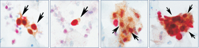
Dual staining positive cells: the cytoplasm stained brown yellow and the nucleus stained red (marked by the arrow)
 p16 & Ki67 cervical cells smear
 p16 & Ki67 cervical tissue section


