
-
-
SHOP

Understanding CISH: Main Advantages and Practical Applications
In the field of pathology, looking at a cell’s setup under a microscope gives useful details. But what if you could observe more? What if you could see the genetic directions and issues occurring inside that same cell, all on one slide? This is the strength of Chromogenic In Situ Hybridization (CISH). It is an innovative method that introduces molecular diagnostics into the regular world of each lab.
This overview examines the basics of CISH, its main benefits over other approaches, and its important uses in clinical diagnostics. At the same time, it shows how this strong method can fit smoothly into your routine.
Understanding CISH: Molecular Diagnostics in a New Light
What is Chromogenic In Situ Hybridization (CISH)?
At its center, CISH is a molecular pathology approach applied to find certain DNA or RNA sequences inside a cell or tissue part. The method employs a labeled DNA probe that attaches to its matching sequence in the cell’s nucleus. Unlike other ways that rely on fluorescence, CISH uses an enzymatic response that creates a colored deposit at the target area. The outcome is a clear, colored spot or mark that can be seen with a usual bright-field microscope. This is the same microscope used for standard H&E staining.
CISH vs. FISH: The Critical Bright-Field Advantage
Many labs know Fluorescence In Situ Hybridization (FISH), a comparable method that applies fluorescent probes. While effective, FISH needs a special, costly fluorescence microscope and skilled staff. In addition, the fluorescent marks can weaken over time, which makes storage hard.
CISH handles these challenges. By generating a lasting, chromogenic mark, it lets pathologists do several things. They can apply current bright-field microscopes. They can review genetic marks and tissue structure at the same time. They can store slides for ongoing review and discussion.
The Main Advantages of Integrating CISH into Your Lab
Accessibility and Cost-Effectiveness
The option to use regular lab gear makes CISH a very reachable and budget-friendly starting point for molecular diagnostics. There is no requirement for spending on pricey fluorescence tools or darkroom setups.
Simultaneous Analysis of Tissue Morphology
One of the biggest benefits of CISH is the preservation of the tissue background. The pathologist can assess the genetic mark, such as gene increase, while viewing the nearby cellular layout. This combined sight is vital for issuing a correct and full diagnosis. Some research has indicated strong agreement rates between CISH and FISH for main biomarkers. This backs CISH’s trustworthiness while adding a structural background. This detail comes from outside the given sources.
Permanent Results for Archiving and Review
The chromogenic marks produced in a CISH test are steady and do not weaken. This permits glass slides to be stored without limit, which is necessary for past studies, quality checks, and sharing cases with coworkers.
Practical Applications: Where CISH Makes a Difference
CISH is not only a conceptual tool. Instead, it holds deep real uses in identifying and handling diseases.
Detecting Viral Infections
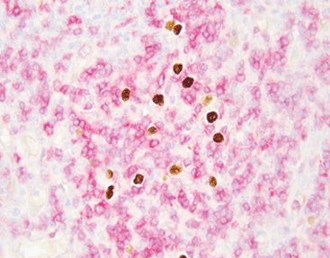
CISH works well for spotting hidden viral infections within tissue. For instance, finding the Epstein-Barr virus (EBV) is key in identifying specific lymphomas and nasopharyngeal carcinomas.
Assessing Gene Amplification in Cancer
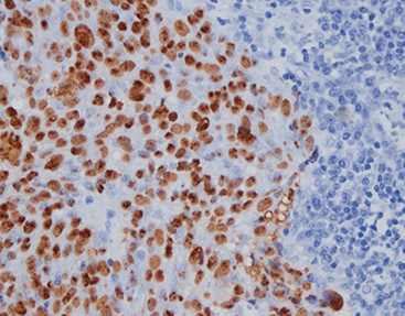
In oncology, CISH is applied to spot the increase of genes like HER2 in breast and gastric cancer. A raised number of gene copies, shown as several colored spots, can direct oncologists in choosing targeted treatments.
Characterizing Hematological Malignancies
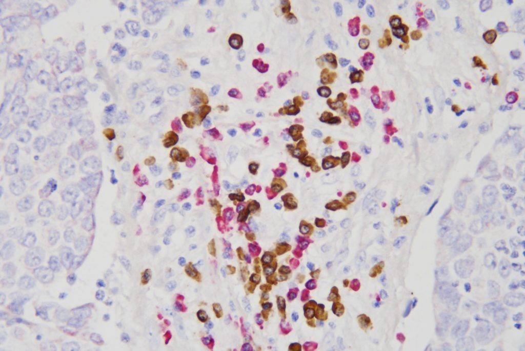
In hematopathology, figuring out the clonality of B-cell groups is crucial to identifying lymphomas. Probes for Kappa and Lambda light chains can assist in spotting a monoclonal group, which confirms a malignancy.
Celnovte: Powering Your CISH Workflow with Precision Tools
At Celnovte, we commit to supplying labs with the sturdy and dependable tools required to carry out advanced diagnostics with assurance. Our molecular pathology collection is crafted to bring clarity and speed to your CISH routine.
High-Sensitivity CISH Probes for Confident Diagnosis
The correctness of any CISH test relies on the standard of the probe. We provide a selection of high-performance CISH and ISH probe kits built for remarkable detail and signal strength. Our products include:
• EBER Probe (Automated & Manual): Tailored for the spotting of Epstein-Barr virus, our EBER probe serves as a vital tool in cancer and infectious disease pathology. We supply versions adjusted for both automated and manual steps to match your lab’s requirements.
• Kappa/Lambda Probe Kit (ISH): This kit is necessary for hematopathology labs to evaluate B-cell clonality in lymphoma diagnostics. It offers clear, simple-to-read outcomes.
• EBER/IHC Dual Staining Kit: Advancing further, this modern kit permits the combined spotting of EBV (with an ISH probe) and a protein marker (with an IHC antibody) on the same slide. As a result, it delivers a lot of details from one tissue part.
Achieving Consistency with the Automated ISH Processor
Repeatability is central in a clinical lab. Our automated ISH Processor is designed to simplify the whole in situ hybridization process, from baking to staining. By automating these involved and lengthy steps, the ISH Processor does a few things. It cuts down on direct time and lowers the chance of human mistakes. It secures standard processing settings for each slide. It provides uniform, top-quality outcomes every time.
When you pair our adjusted CISH probes with the accuracy of our ISH Processor, you form a strong, trustworthy, and quick system for molecular diagnostics.
Conclusion
Chromogenic In Situ Hybridization connects the divide between usual histology and molecular genetics. It presents a strong, reachable, and insightful diagnostic approach. This enables labs to find key details that direct patient care without needing large expenditures on special gear.
Celnovte acts as your perfect ally in applying and improving this technology. With our better CISH probes and modern automated ISH Processor, you can introduce the future of molecular pathology into your lab now.
FAQ
Q: What is the main difference between CISH and FISH?
A: The main difference is the detection method. CISH uses an enzyme to create a colored signal visible with a standard bright-field microscope, while FISH uses fluorescent molecules that require a special fluorescence microscope.
Q: Can CISH be automated?
A: Yes. The CISH procedure can be fully automated using an instrument like the Celnovte ISH Processor, which improves consistency, reduces turnaround time, and minimizes manual errors.
Q: What is dual staining, like in the EBER/IHC kit?
A: Dual staining allows for the detection of two different types of molecules on one tissue slide. The Celnovte EBER/IHC kit, for example, lets you visualize EBV genetic material (via ISH) and a specific protein (via IHC) at the same time.
Q: Are CISH results permanent?
A: Yes. The chromogenic precipitate that forms the signal in CISH is stable and does not fade, allowing slides to be archived for long-term storage and future review.