 PRODUCT CATEGORY
CONTACT US
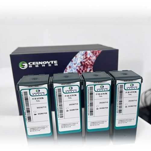
MMR panel | MLH1 (C12A19), MMab
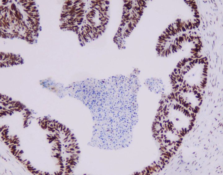
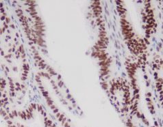
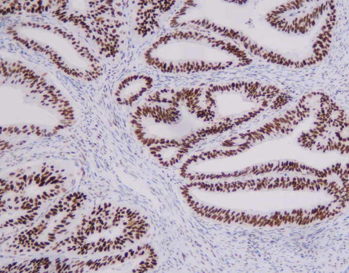
MLH1 is a mismatched fix protein in molecular weight of about 87kDa size which is usually associated with the occurrence of hereditary non-polypolycinary colon cancer. The functional defects of MLH1 are considered to be an important mechanism for tumor morbidity and are used to determine genetic mutations in colorectal, esophageal, stomach and cervical cancers. The study showed that about 20% of colorectal cancer patients had a mismatch repair protein (MMR) of expression deficiency, while in the distributive colorectal cancer , MLH1 deficiency patients accounted for more than 95% of patients with MMR deficiency.
More Info
[Intended Use]
This antibody is intended for use in Immunohistochemical applications on formalin-fixed paraffin-embedded tissues (FFPE). The clinical interpretation of any staining or its absence should be complemented by morphological studies using proper controls and should be evaluated within the context of the patient’s clinical history and other diagnostic tests by a qualified pathologist.
[Specifications]
| Product Name | MLH1 (C12A19), MMab |
| Catalog No. | CMM-0184 |
| Intended Use | IVD, RUO |
| Species Reactivity | Human; others not tested |
| Cellular Localization | Nuclear |
| Antibody Type | Mouse Monoclonal |
| Clone | C12A19 |
| Format and Volume | Ready-to-use: 1.5mL, 7.5mL Concentrated: 0.1mL, 0.2mL and 1mL |
[Datasheets & SDS]
| IVD Datasheet (IFU) | ↕️ Download |
| RUO Datasheet (IFU) | ↕️ Download |
| SDS sheet | check with sales |
[Storage and Validity]
Store at 2~8°C. Avoid freezing.
Maintain temperature below room temperature during transport, ensuring it does not exceed one week.
[Protocol Recommendations]
This reagent has been optimized on the fully automatic immunohistochemical stainer and does not require reconstitution, mixing, dilution or titration.
For detailed operating parameters, please refer to the operating manual of the fully automatic immunohistochemical stainer.
1.Test Procedure:
1.1 Incubation with peroxidase block quenches the activity of endogenous peroxidase.
1.2 Use MSH6 to incubate.
1.3 Linker , rabbit-anti-mouse incubation.
1.4 Use polymer for incubation, and bind the reagent to linker , rabbit-anti-mouse .
1.5 Incubate the antigen-antibody complex with DAB, and finally appear in the form of a brown precipitate, which can be observed under a light microscope.
1.6 Use DAB Enhancer for incubation to enhance color intensity.
1.7 Incubate with hematoxylin and counterstain the sections.
1.8 Use bluing reagent to incubate and blue hematoxylin to enhance the contrast of the staining results.
2.Quality Control
2.1 Positive Control
Positive controls serve as an indication of correct tissue preparation and appropriate staining techniques. Each staining should include a positive pair of photos for comparison under the same test conditions. Known positive tissue controls should only be used to monitor the correct performance of procedures and testing of reagents and are not intended to aid in describing a definitive diagnosis of a patient sample. If the positive tissue control fails to show appropriate positive staining, the results of this batch of experimental test samples should be considered invalid.
2.2 Blank Control
Each staining should include a blank control reagent under the same test conditions for comparison. Blank control reagents are used to stain tissue sections instead of antibodies to judge non-specific staining and provide a better explanation for antigen site-specific staining.
[Reference]
1. Shia J. J Mol Diagn. 2008;10(4):293–300.
2. Bellizzi AM. Surg Pathol Clin. 2017;10(4):843–868.
3. Park JY et al. Pathol Res Pract. 2012;208(7):428–433.
4. Hendricks LA et al. J Pathol. 2004;204(1):28–35.
Related Product


 PRODUCT CATEGORY



 Chat
Chat

 message

 Quote
Quote

