
The Path to Precision in Cytopathological Diagnosis
Immunocytochemistry has become a routine procedure in cytopathology.
Advantages of Immunocytochemistry Technology
High sensitivity and strong specificity
Provides reliable diagnostic information
Clinical Applications of Immunocytochemistry
Identifying metastatic carcinoma of unknown primary
Differentiating mesothelioma from adenocarcinoma
Distinguishing poorly differentiated carcinoma from lymphoma, among others
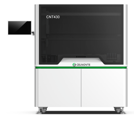
Direct Loading of Cell Samples--Fully Automated Immunocytochemistry Staining System
Suitable for both gynecological and non-gynecological cytopathological samples.
From Slide Preparation to Staining – Morphology & Immunocytochemistry – All Done on One Machine
Load samples and start with one click for both morphological and ICC staining.
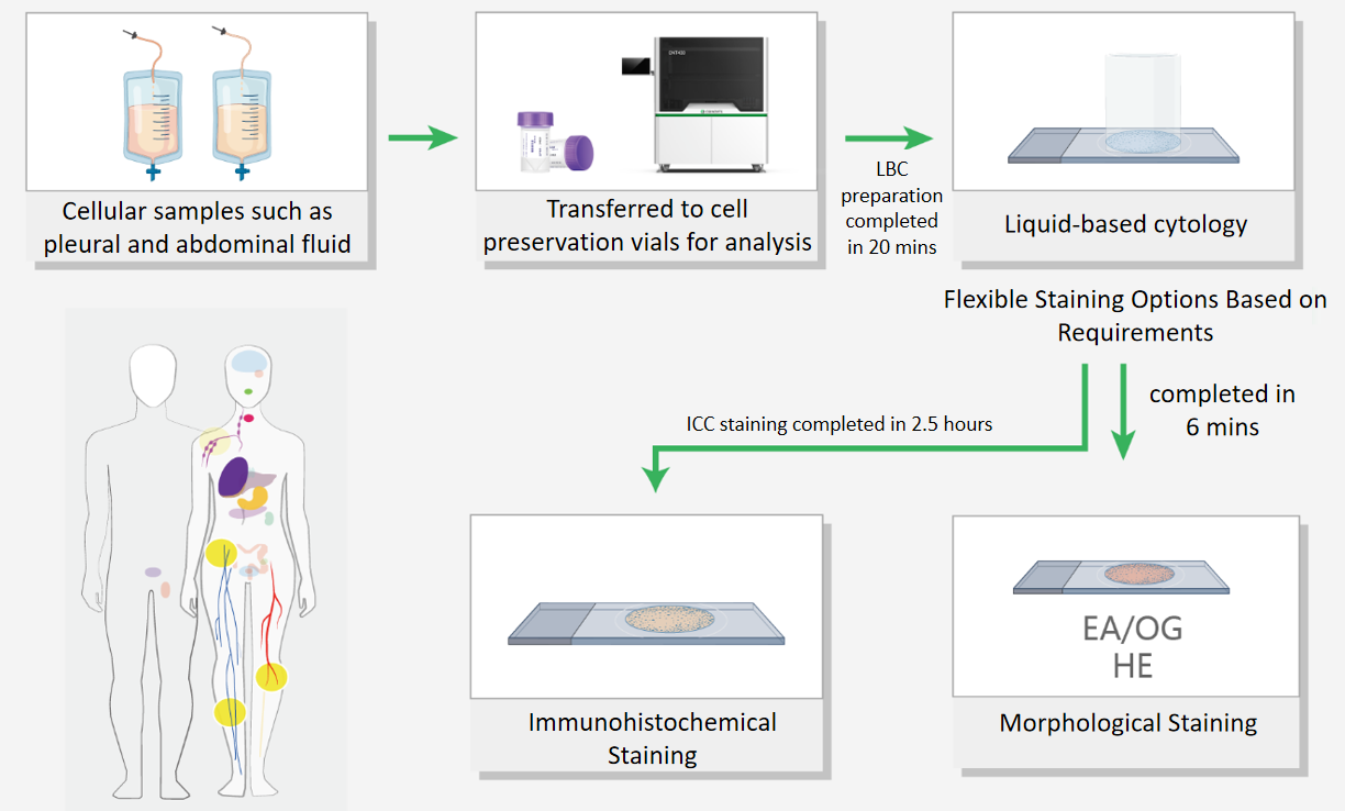
LBC Preparation: Efficient and Rapid
Real-Time Standardized Immunocytochemistry Testing
Recommended by CAP (College of American Pathologists) guidelines--Suitable for minute cell specimens, enriching more cells.

Real-time testing: Obtain staining results promptly.
Broad sample applicability: Suitable for minute specimens and samples difficult to embed in paraffin blocks.
Clean background: Especially improves results for hemorrhagic serous effusion specimens.
Reliable results: Enriches a higher number of cells.
Repeatable slide preparation: Allows preparation of multiple LBC slides for staining.
Simple and efficient: Avoids human errors associated with paraffin block preparation.
Dual-Chamber Single Slide, Flexible Selection – Morphological Staining + Immunomarkers
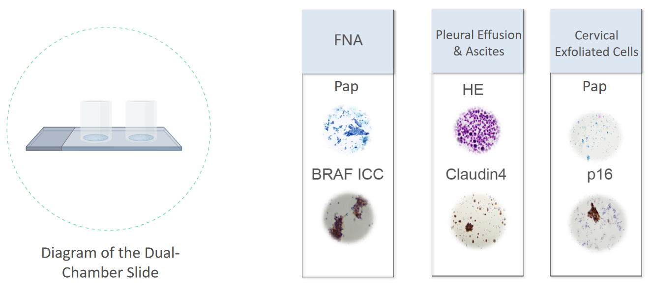
Gynecological Staining Panel
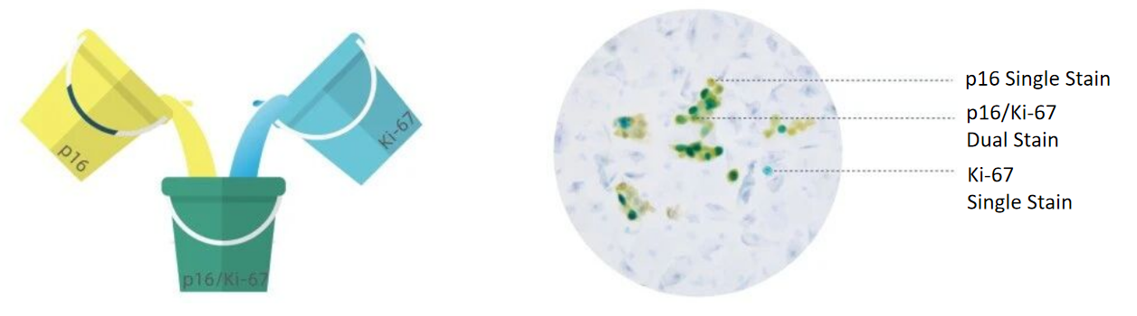
p16/Ki-67 Cytology Dual Staining
Dual chromogenic system for flexible options
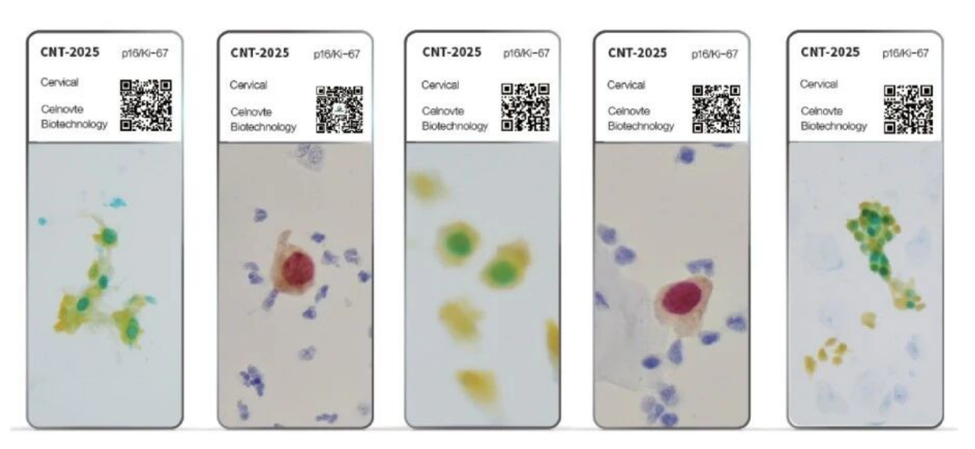
Easier-to-Interpret Chromogen and Color System Combination
Yellow (p16) + Indigo (Ki-67) = Overlapping Green (Dual-Stain Positive)
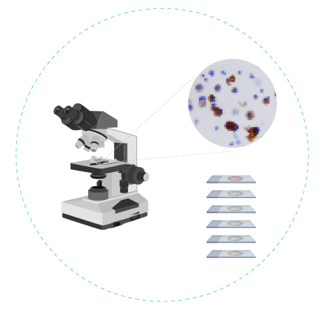
Under the microscope, distinct "green dots" are clearly visible at a glance, effectively solving interpretation challenges.