


Trichrome-Masson Stain Kit
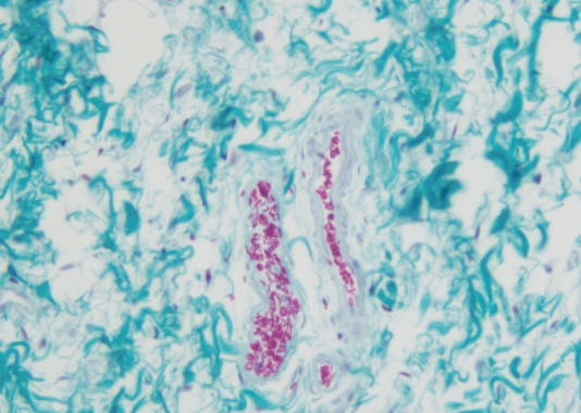
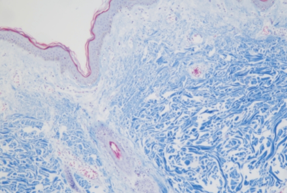
It is mainly used to distinguish collagen fibers and myofibrils, and can also be used for renal biopsy specimens. Collagen fibers are green or blue, and myofibrils are red. It is also important for judging the precipitation of immune complexes.
Product Features
Product Application:
Clearly display the distribution of collagen fibers and muscle fibers in liver tissues, for the diagnosis of hepatic fibrosis, renal interstitial fibrosis, myocardial fibrosis and other diseases.
Staining Principle:
Tissue components differ in density, leading to varied capacity for adsorbing and retaining dye molecules.
Staining Results:
Nuclei appear blue‑black; collagen fibers are blue; muscle fibers are red.
Product Specifications
| Product Category | Cat.No. | Description | Specification |
|---|---|---|---|
| Manual Reagents | HS2001 | Aniline Blue Method‑Manual | 4×5 mL/kit4×10 mL/kit4×20 mL/kit4×50 mL/kit4×100 mL/kit |
| Manual Reagents | HS2002 | Light Green Method‑Manual | 4×5 mL/kit4×10 mL/kit4×20 mL/kit4×50 mL/kit4×100 mL/kit |
| CNT520 On‑board Reagents | HS2211 | Aniline Blue Counter‑stain Type | 30 tests/kit |
| CNT520 On‑board Reagents | HS2212 | Light Green Counter‑stain Type | 30 tests/kit |
-
Product Features
-
Product Specifications