

Reticular fiber stain kit
The histological changes of reticular fibres are of great importance in the pathological diagnosis of clinical diseases, such as haematological tumours, breast fibroepithelial tumours, gastrointestinal mesothelioma and neurological tumours. It is important to observe the number, thickness, density, breakage and collapse of reticulocytes in the lesion through reticulocyte staining to determine the nature, extent, development and regression of the lesion.
Product Features
Product Application:
To evaluate the degree of liver fibrosis, differentiate carcinoma in situ from invasive carcinoma, diagnose myelofibrosis, and perform differential diagnosis of tumors.
Staining Principle:
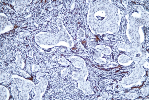
Tissues are oxidized by potassium permanganate to expose aldehyde groups, which specifically adsorb silver ions and turn black after reduction by formaldehyde.
Staining Results:
Reticular fibers appear black.
Specifications
| Product Category | Cat.No. | English Name | Registration Certificate Name | Description | Specification |
|---|---|---|---|---|---|
| Manual Reagents | HS2010 | Reticular fiber stain kit | Reticular Fiber Staining Solution | Reticulum‑silver Staining | 7×5 mL/kit7×10 mL/kit7×20 mL/kit7×50 mL/kit7×100 mL/kit |
| CNT520 On‑board Reagents | HS2205 | Reticular fiber stain kit | Reticular Fiber Staining Solution | — | 30 tests/kit |
-
Product Features
-
Specifications