


Why Primary Antibody Compatibility Matters in IHC Staining
Reliable immunohistochemistry staining depends on more than primary antibody specificity. Tissue preparation, antigen retrieval, antibody incubation, detection chemistry, and washing conditions all influence the final staining result. Even when a primary antibody performs well under manual conditions, its performance may change when transferred to an automated IHC stainer.
For clinical pathology laboratories, the goal is to establish a workflow that supports specific staining, appropriate signal intensity, low background, and reproducible results across slides and staining runs.
Matching an IHC staining platform to primary antibody requirements therefore involves evaluating both the antibody's established protocol and the instrument's capabilities. Rather than changing several parameters simultaneously, laboratories should identify which conditions must be preserved and which can be standardized through automation.
Identify the Primary Antibody's Critical Requirements
Before introducing an antibody into an automated workflow, review its validated or established staining protocol. Different antibodies may require different antigen retrieval methods, incubation periods, working concentrations, and detection systems.
Three areas deserve particular attention:
Antigen retrieval: Heat-induced epitope retrieval conditions, including buffer selection, pH, temperature, and duration, can influence epitope accessibility. Conditions suitable for one antibody may not produce the same results with another.
Primary antibody incubation: Concentration and incubation time affect the interaction between the antibody and its target. Insufficient incubation may weaken the signal, while excessive concentration or incubation may increase nonspecific background.
Detection compatibility: The secondary antibody or polymer detection system must be compatible with the primary antibody's host species and immunoglobulin characteristics. The detection chemistry must also match the intended visualization method.
These requirements provide a practical starting point for evaluating an automated platform. The objective is not to make every antibody follow an identical protocol, but to determine whether the instrument can accommodate the conditions necessary for each assay.
Evaluate How an Automated IHC Stainer Handles Different Conditions
An automated IHC stainer can standardize reagent application, incubation timing, washing, and other programmed steps. However, automation alone does not guarantee compatibility with every primary antibody.
When evaluating a platform, laboratories should examine the following capabilities:
Flexible protocol settings. Can the instrument accommodate different incubation times and staining sequences? Flexibility is particularly important when laboratories process a broad antibody panel with different performance requirements.
Consistent reagent delivery. Uniform reagent coverage helps reduce variation caused by uneven application or tissue drying. Laboratories should also consider how the system manages washing and reagent removal.
Control of incubation conditions. Stable and repeatable processing conditions help laboratories reduce operator-dependent variation between runs.
Detection-system compatibility. The platform should support the intended detection workflow, subject to the manufacturer's specifications and the laboratory's validated procedures.
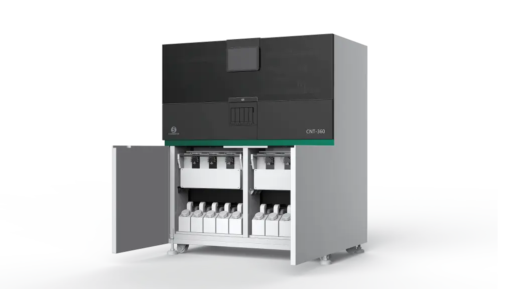
Celnovte Biotech offers automated immunohistochemistry platforms and integrated pathology solutions for laboratories evaluating these workflow requirements. Its CNT360 Full Automatic IHC&ISH Stainer is one option to examine when assessing automated staining capabilities.
Explore the CNT360 automated IHC stainer to evaluate how automated processing can support standardized immunohistochemistry workflows.
Optimize Staining Conditions Without Losing Antibody Performance
Moving from manual to automated immunohistochemistry staining should be treated as a method-transfer exercise rather than a direct replacement of manual steps.
Start with the existing protocol and identify the variables most likely to affect staining performance.
Antigen Retrieval Conditions
Review the retrieval buffer, pH, heating conditions, and cooling procedure. These parameters can influence both signal strength and tissue morphology. If the automated platform uses a different retrieval process, compare the resulting staining patterns against appropriate controls before routine implementation.
Primary Antibody Incubation
Establish whether the current antibody concentration and incubation period can be maintained on the instrument. If adjustments are necessary, change one variable at a time and document the effect. Avoid assuming that a longer incubation or higher concentration will always improve staining.
Washing and Background Control
Insufficient washing may contribute to nonspecific staining, while inappropriate processing can affect tissue integrity or signal retention. Consistent washing conditions, adequate reagent removal, and prevention of tissue drying are therefore important when transferring protocols.
Detection and Signal Development
The detection system, enzyme activity, and chromogen development conditions influence the final signal. A change in detection chemistry may require reassessment of the complete staining workflow rather than an isolated adjustment to primary antibody incubation.
These parameters should be evaluated together because they interact. Stronger staining does not necessarily indicate better performance if background increases or the expected cellular localization becomes less distinct.
Validate Compatibility Before Routine Use
A practical compatibility assessment should compare the established method with the proposed automated workflow using appropriate tissue controls and documented acceptance criteria.
A structured evaluation can include:
Positive controls: Confirm that the expected target produces an appropriate staining signal and localization.
Negative or blank controls: Assess background staining and potential nonspecific reactions.
Repeatability: Compare replicate sections or repeated runs for differences in intensity, localization, and background.
Tissue integrity: Check whether the processing conditions preserve morphology and permit reliable interpretation.
Protocol documentation: Record retrieval conditions, antibody concentration, incubation time, detection reagents, and any instrument-specific adjustments.
When results differ from the established method, investigate the source of variation before changing multiple parameters. Tissue fixation, section preparation, antibody handling, and detection chemistry can all contribute to differences.
Acceptance criteria should be defined according to the assay's intended use and applicable laboratory procedures. Automated staining should not be considered validated simply because the instrument completes the programmed sequence.
Match the Platform to the Laboratory's Workflow
The most suitable IHC stainer depends on more than antibody compatibility. Laboratories should also consider daily slide volume, the number of antibodies in routine use, the need for multiplex workflows, and future automation requirements.
For routine single-target staining, prioritize reliable execution of established protocols and manageable workflow standardization. Laboratories handling more complex staining requirements should also assess whether the platform supports their intended multiplex applications and associated reagent workflows.
Celnovte Biotech provides several platform options for laboratories with different automation needs:
The CNT300 Full Automatic Multiplex IHC Stainer is worth evaluating for laboratories considering multiplex IHC workflows.
The CNT320 Fully Automated IHC/ISH Stainer provides another option for laboratories assessing automated IHC and ISH processing.
The CNT330 Full Automatic Multiplex IHC Stainer is another platform to consider when evaluating multiplex staining requirements.
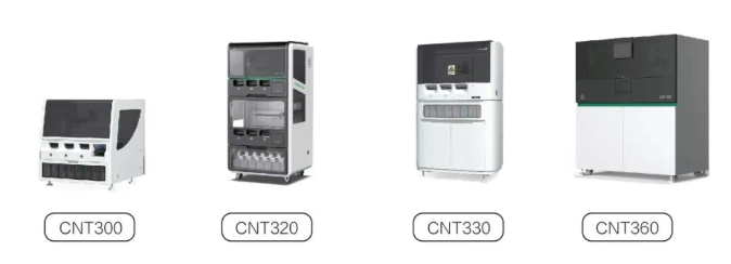
Instrument specifications, supported protocols, reagent compatibility, and throughput should be confirmed with the manufacturer against the laboratory's actual requirements.
For a broader view of instrument and reagent integration, see Celnovte Biotech's Fully Automated Immunohistochemistry Complete Solution. It provides an overview of the company's integrated approach to automated IHC instruments and immunostaining reagents.
Conclusion
Successful immunohistochemistry staining requires a coordinated relationship between primary antibody characteristics, tissue preparation, retrieval conditions, incubation parameters, and detection chemistry. An automated IHC stainer can improve procedural consistency, but each assay still requires appropriate compatibility assessment and validation.
For pathology laboratories selecting an automation platform, the most effective approach is to document critical antibody requirements, evaluate the instrument's protocol flexibility, compare results using appropriate controls, and establish reproducible operating conditions before routine use.
By treating antibody compatibility as a complete workflow consideration, laboratories can make more informed IHC platform decisions while maintaining the quality and interpretability of staining results.